Quiz

Quiz Aug/Sep 2025
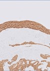
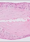
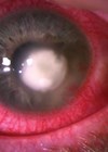
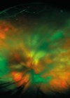
History A 78-year-old female presented with bilateral, painless, progressive blurring of vision over five years, photophobia and increasing glare. Her past medical history included a known diagnosis of monoclonal gammopathy of undetermined significance (MGUS). On examination, vision in the left...

Quiz Oct/Nov 2024
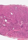
History An 83-year-old female was previously treated by surgical excision and plaque brachytherapy for her left conjunctival lesion. She presented to her ophthalmologist with a recurrence some years later and underwent a lid sparing orbital exenteration, which was sent to...

Quiz Jun/Jul 2024
History A one-year-old girl originally presented with a red eye, initially bilaterally and later in the left only. On examination a membrane was found on the tarsal conjunctiva of the upper eyelid (Figure 1). Figure 1: Anterior segment. Serum plasminogen...
Quiz Feb/Mar 2024
History A 21-month-old boy presented to his local ophthalmology department with a left proptotic eye from a growing cystic lesion known to be present from birth. Notes taken on presentation were: Known left microphthalmia with chorio-retinal coloboma, contralateral eye was...
Quiz Apr/May 2023
History A 76-year-old female presented at her local district general hospital with right decreased visual acuity, glare, and foreign body sensation, in addition to longer-term dry eyes. She was otherwise well. Her past medical history included hypertension. On examination: vision...
Quiz Dec/Jan 2023
History A 35-year-old female presented to the emergency eye clinic with an acutely red, painful, photophobic left eye. She was a contact lens-wearer but denied swimming, showering, or sleeping in her lenses. She resided on a farm and worked as...

Quiz Oct/Nov 2022
History An Afro-Caribbean woman, aged 30, with “scleral tattooing” for cosmetic reasons (subconjunctival dye injection), presented with bilateral upper and lower eyelid oedema for one week, and she also had restricted eye movement and blurry vision in her right eye....

Quiz Aug/Sep 2022
History A 58-year-old female patient was referred with a 2-year history, gradually enlarging, painless left upper lid lump. There was no bleeding or ulceration. She had no other eye symptoms and no other skin lesions. Her past medical history included:...
Quiz Feb/Mar 2020
History A 92-year-old female patient was referred for a three-month history of a left conjunctival growth with ocular irritation. Her past medical history included: hypertension, back pain and osteoarthritis, all of which were controlled by medication. On examination: vision in...
Quiz Dec/Jan 2020
History A 69-year-old female patient was referred to the uveitis clinic from her local district general hospital with a left posterior uveitis which had been unresponsive to high dose steroids. She had no other previous ophthalmic history nor significant systems...
Oct/Nov 2019 Quiz
History A 62-year-old female patient was referred for rapid growth of a left periorbital soft tissue lesion with proptosis. Her past medical history included: hypertension, hyperlipidaemia and bipolar disorder. On examination: vision in the affected eye was hand movements, right...
Aug/Sep 2019 Quiz
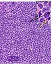
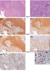
History A 73-year-old female patient was referred for rapid growth of two new lesions on the face. She had past medical history of systemic lymphoma. On examination, there was a palpable mass over the left superior orbital region and the...