History
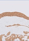
A one-year-old girl originally presented with a red eye, initially bilaterally and later in the left only. On examination a membrane was found on the tarsal conjunctiva of the upper eyelid (Figure 1).

Figure 1: Anterior segment.
Serum plasminogen level was 28% and she was being treated with serum eyedrops and topical steroids. Multiple excisions had been undertaken with the latest one (at age four) being sent to the ophthalmic pathology department. Six weeks after excision, a small flat recurrence was noted. There was no other significant past medical history of note, in particular involvement of other mucous membranes was not reported.

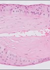
Figure 2: H+E 2x.

Figure 3: H+E 20x.
Questions
Figures 2 and 3 show representative H+E-stained sections of the lesion. Special stain Congo red was also done but this was negative (not shown).
- How can this be described?
- Considering the clinicopathological features, what is the diagnosis?
- What is the potential histological differential diagnosis prompting Congo red staining?
- What are the management options?
Answers
- Figures 1 and 2 demonstrate ulcerated conjunctiva with active chronic inflammation, granulation tissue and thick and fibrinous dense eosinophilic amorphous hyalinised material. The eosinophilic deposits were negative for Congo red stain.
- he overall features are those of ligneous conjunctivitis. This is a rare, chronic, recurrent conjunctivitis characterised by fibrinous ‘wood-like’ pseudomembranes on the palpebral conjunctiva. The pseudomembranes often involve the upper tarsal conjunctiva, although the condition may also involve the bulbar and lower conjunctiva, and typically separate without bleeding. Patients have a history of recurrent conjunctivitis in response to external irritants, fever, trauma (including surgery) or treatment with antifibrinolytic drugs. Similar pseudomembranous lesions may be observed in other mucous membranes of the oral, respiratory, gastrointestinal and female genital tract, but lesions have also been reported in the ear and renal collecting system.Ligneous conjunctivitis is usually diagnosed in young children, although it can present at any age with a slight female preponderance. It can occur sporadically or inherited as an autosomal recessive trait by mutations in the plasminogen type-1 gene (on chromosome 6q26–27) resulting in plasminogen deficiency (hypoplasminogenemia). Decreased plasminogen activity leads to impaired wound healing with lack of plasmin-mediated extracellular fibrinolysis and subsequent development of fibrin-rich deposits. Histomorphologically, the pseudomembranes are stromal deposits of amorphous eosinophilic material (fibrinogen) with granulation tissue and inflammatory cell infiltrate.
- The histological differential diagnosis to exclude is amyloidosis, which would stain positively with Congo red with characteristic ‘apple-green’ birefringence under polarised light.
- Management usually consists of a combination of surgical and medical therapies to prevent complications and preserve ocular health, but the lesions do recur and multiple / ongoing treatments are likely to be needed. Surgical removal of the lesions is usually necessary, but the trauma can result in rapid recurrence; combining with medical therapy is more likely to control recurrence or at least slow it down. Amniotic membrane transplantation following surgical excision of the pseudomembranes has also been shown to be effective. Medical options include topical and systemic fresh frozen plasma, topical plasminogen, topical heparin, topical corticosteroids and topical cyclosporine A.
COMMENTS ARE WELCOME