History
An 83-year-old female was previously treated by surgical excision and plaque brachytherapy for her left conjunctival lesion. She presented to her ophthalmologist with a recurrence some years later and underwent a lid sparing orbital exenteration, which was sent to the ophthalmic pathology department. There was no other significant past medical history of note.
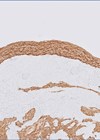
The orbital exenteration specimen comprised the upper and lower lids, globe, optic nerve stump and surrounding orbital soft tissue (Figure 1). A partially pigmented inferior bulbar conjunctival mass was noted.

Figure 1.

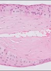
Figure 2a: H&E x20.

Figure 2b: MelanA x20.
Questions
Figure 2 shows a representative H+E and a melanA immunohistochemical stained section of the lesion.
- How can this be described?
- Considering the clinicopathological features, what is the diagnosis?
Answers
- Figures 1 and 2 demonstrate a large, nodular partially pigmented invasive melanoma of the inferior bulbar conjunctiva. The melanoma cells were predominantly of epithelioid cell type and were strongly immunopositive for melanA. There was a mild, patchy infiltration of tumour infiltrating lymphocytes and melanophages. Other features present but not shown here, include: overlying conjunctival melanoma in situ; perineural invasion; and involvement of the trabecular meshwork by melanoma cells. The melanoma was 1.5mm from the deep cutaneous surgical margin of the lower lid. All other surgical margins were clear. The inferior forniceal and palpebral conjunctiva were clear. Likewise, the lower eyelid skin, upper eyelid skin and conjunctiva, lacrimal gland, nasolacrimal system and optic nerve were all clear of in situ and invasive melanoma. There was no BRAF V600E mutation present.
- This was a recurrence of invasive conjunctival melanoma with intraocular and orbital involvement. Conjunctival melanoma is a rare, aggressive invasive ocular surface tumour with an overall incidence of 0.46 cases per million persons per year, more common in those over 65 years of age and predominantly affects fair complexioned populations. It represents around 0.25% of melanomas at all sites and 5% of all ocular melanoma and is the second most common conjunctival malignancy after squamous cell carcinoma.
It is often unilateral and can affect any part of the conjunctiva but commonly presents on the bulbar surface, near the limbus. Invasion of the cornea, eyelid, sclera or orbit may occur in advanced tumours.
Conjunctival melanoma is a mucosal melanoma with similar histological and UV-related molecular signatures (BRAF, NF1 and RAS gene mutations) to cutaneous melanoma. The tumour originates from the basal melanocytes in the conjunctival epithelium. The majority (~70%) of cases develop from conjunctival melanocytic intraepithelial lesions (C-MIL) / primary acquired melanosis with atypia (PAM), while others arise from pre-existing nevi or de novo.
There is no standard of care for C-MIL or conjunctival melanoma; consequently, management varies considerably between ophthalmic and specialised ocular oncology centres. This includes wide local surgical excision +/- amniotic membrane allograft and +/- adjuvant cryotherapy, topical chemotherapy (mitomycin C, 5-fluorouracil or interferon alpha-2b), radiotherapy (brachytherapy, proton beam or photon external beam), or radical orbital exenteration for advanced cases with local tissue invasion (as in the case presented here).
Postoperative complications rates and risk of tumour recurrence are high (33–61%), warranting long-term follow-up. Poor prognostic indicators / risk factors for nodal (~25%) and systemic metastases include a non-epibulbar locations, ulceration and increased tumour thickness.