Top Tips
Headaches in ophthalmology (part 2)
Ophthalmologists see a large number of patients with headaches or facial pain in the ophthalmic outpatient clinics or in emergency clinics. Over two articles, I will discuss several causes of headaches, ocular manifestations and proposed management and referral options. It...
Headaches in ophthalmology (part 1)
Ophthalmologists see a large number of patients with headaches or facial pain in the ophthalmic outpatient clinics or in emergency clinics. Over two articles, I will discuss several causes of headaches, ocular manifestations and proposed management and referral options. It...
What not to miss in neuro-ophthalmology Part 2
As mentioned previously there are several conditions in neuro-ophthalmology that should not be missed by the general ophthalmologist as well as ophthalmology trainees. We discussed in the first part some of these conditions including third cranial nerve palsies, giant cell...
What not to miss in neuro-ophthalmology Part 1
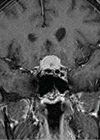
Neuro-ophthalmology is a complex and difficult subspecialty in ophthalmology. It has several connections to neurology, neuro-surgery, rheumatology as well as many other medical specialties. Working in an multidisciplinary team (MDT) environment is key to success in this subspecialty as mistakes...
A practical guide to anisocoria
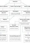
Anisocoria means the presence of difference in the size of the right and left pupils. It is a sign of an abnormality in the efferent pathway. The first question facing the ophthalmologist is to ascertain if anisocoria is present or...
Vitreomacular traction and full thickness macular hole
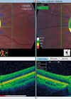
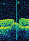
Clinical scenario: A 64-year-old lady presented to the clinic with a few weeks history of sudden onset of metamorphopsia, central blur and reduced vision in her right eye. The ocular examination and ocular coherence tomography confirmed right eye focal vitreomacular...
Diagnosis and management of IV cranial nerve palsy
Aetiology: Trochlear nerve palsy can be divided into acute or congenital. Congenital trochlear nerve palsy is usually noted in childhood with development of abnormal head posture. Various pathologies can lead to acute IV nerve palsy, most commonly trauma. Other causes...
Third nerve palsy
Case scenarios A 71-year-old female presented to a nearby eye emergency unit with two days history of partial ptosis in her left eye with diplopia. She saw her GP earlier that day and he asked her to go to the...
21st Century retinal laser treatment in the anti-VEGF era
In today’s world, macular laser treatment has a vital role in the treatment of diabetic macular oedema (DMO). DMO is one of the most common causes of visual impairment. Despite expensive intravitreal treatment courses of anti-VEGF, many will agree that...
Strabismus in thyroid eye disease
Pathogenesis Thyroid eye disease (TED) is an auto-immune condition, in the initial phase there is lymphocytic infiltration and oedema of the extraocular muscles with deposition of glycosaminoglycans and hyaluronic acid and adipogenesis, which can lead to an increase in the...
The management of chronic central serous chorioretinopathy
Central serous chorioretinopathy (CSCR) is a common retinal disease characterised by one or more serous neurosensory detachments. Patients present with acute onset blurring of vision, metamorphopsia and / or central scotomas. The condition is six times more common in men...
Ultrasound biomicroscopy (part 2): primary angle closure
Patients with primary angle closure or primary angle closure glaucoma [PAC(G)] comprise a significant subgroup affecting around 10% of glaucoma patients amongst Caucasians. Assessment of the patient with angle closure, or narrow angles, requires gonioscopy. However, whilst identifying the presence...


