
Case Reports

In the blink of an eye
In this case report series, the author examines the incidence of ocular injuries sustained during game shooting in Scotland between 2012–2018. Precise figures for ocular injuries sustained during game shooting are not available. According to Police Scotland, there were 43,790...
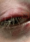
Hidden eyelid laceration following blunt trauma
A paediatric case report of a hidden eyelid laceration following blunt trauma. Blunt injury to the eyelid can result in a multitude of issues, such as damage to the eyelid margin, lacrimal system and surrounding orbit [1]. These can often...
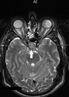
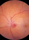
Photophobia: an unusual symptom of a pituitary macroadenoma
Introduction Photophobia, defined as ‘an abnormal intolerance to light’, is commonly associated with a range of both ocular and neurological pathologies such as dry eye, blepharospasm, corneal pathologies, cataracts, uveitis, retinal dystrophies, optic neuritis, migraine, meningitis, and traumatic brain injury...
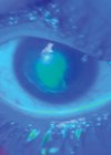
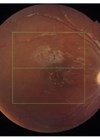
A unique case of macular burn from ‘toy’ laser
The first laser was created in 1960 and its name is an acronym for ‘light amplification by stimulated emission of radiation’. Laser technology has been used for medical, industrial, research and entertainment purposes in a variety of fields following extensive...
Acute dacryoadenitis secondary to COVID-19
Acute dacryoadenitis is defined as the rapid onset of discomfort and swelling of the lacrimal gland, classically giving rise to an S-shaped ptosis [1]. Dacryoadenitis is the most common cause of a painful mass in the lacrimal gland in young...
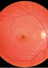
A case of ‘60-day glaucoma’
Neovascular glaucoma (NVG) has been called ‘90-’ or ‘100-day glaucoma’ in the past due to its typical development three months after the onset of central retinal vein occlusion (CRVO). In reality, NVG can occur anywhere between two weeks and two...
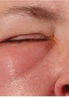
Recurrent unilateral preseptal cellulitis secondary to herpes simplex virus infection
Introduction Periorbital (sometimes called preseptal cellulitis) is a common condition which on its own is not normally an ophthalmic or surgical emergency, however it has the potential to cause severe and serious morbidity in cases where the infection has crossed...
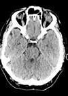
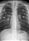
Periorbital and subconjunctival emphysema - a sign of orbital rim fracture
Background Orbital emphysema is a condition where air is present in orbit or periorbital tissues [1]. It is most commonly caused by trauma leading to orbit fracture, where air from paranasal sinuses is allowed to enter the orbit. The most...
Ciliary body granuloma masquerading as a melanoma
Introduction Sarcoidosis is a multisystem granulomatous inflammatory condition which predominantly affects the pulmonary system and intrathoracic lymph nodes, followed by ocular involvement [1]. We present an interesting patient who developed acute anterior uveitis and subsequent ciliary body granuloma clinically mimicking...
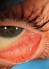
Bilateral eye pain after contact lens wear: an inadvertent case of chemical eye injury
Introduction There are around 4.1 million contact lens wearers in the UK [1]. While the vast majority of them do not experience any complications, over the past years there have been cases of acanthamoeba keratitis and multiple retained contact lenses...

Avoiding investigations through history taking and examinations to differentiate serious from comparably benign aetiology
*Joint first authors. Introduction Anisocoria can be a sign of neurological deficit, necessitating numerous investigations [1]. This case report explores how expensive and time-consuming investigations can be avoided by thorough history taking and examination to differentiate serious from comparably benign...
A paediatric case of central retinal artery occlusion following antibiotics and decompression surgery for orbital cellulitis
Orbital cellulitis is an ophthalmic emergency that warrants urgent management in the hospital setting [1]. This occurs more frequently in the paediatric population where it is often secondary to sinus infections. Delay in treatment could result in severe complications including...

