Introduction
Photophobia, defined as ‘an abnormal intolerance to light’, is commonly associated with a range of both ocular and neurological pathologies such as dry eye, blepharospasm, corneal pathologies, cataracts, uveitis, retinal dystrophies, optic neuritis, migraine, meningitis, and traumatic brain injury [1,2]. Here we describe an unusual case of pituitary macroadenoma presenting with photophobia as an isolated symptom.

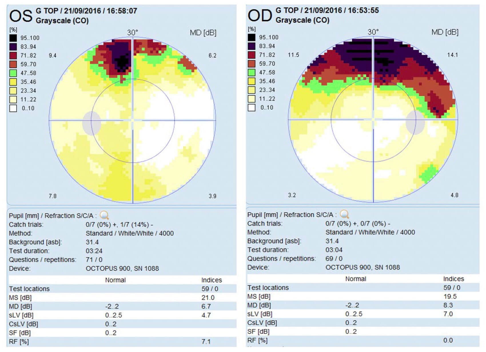
Figure 1: Visual field data demonstrating mild bilateral superior loss which did not respect the midline.
Case
A 40-year-old male presented to the emergency eye clinic with a history of painful sensitivity to light lasting several months, significantly worsening in the previous week. He had no history of neck stiffness, fever, motor or sensory disturbance, or concerning features consistent with raised intracranial pressure. Visual acuity and ocular examination were normal. The anterior chamber was quiet. Visual fields showed mild bilateral superior loss which did not respect the midline (Figure 1). Conjunctival swabs for viral, bacterial and chlamydial infection were negative. Optical coherence tomography of the peripapillary retinal nerve fibre layer was unremarkable. The patient was noted to have unusually large hands, but history and physical examination for other signs of acromegaly were unremarkable. He was therefore managed supportively with ocular lubricants.

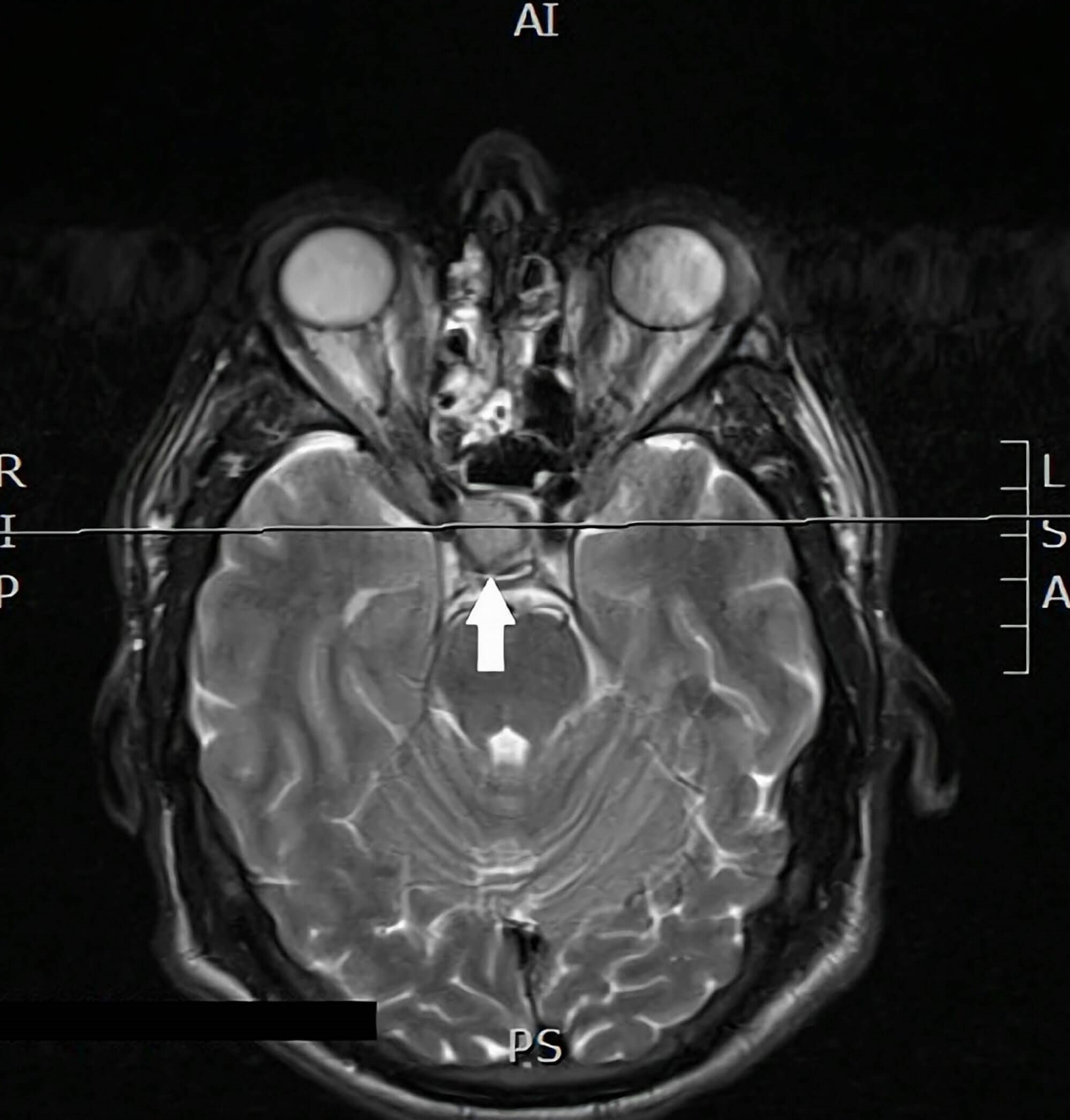
Figure 2: MRI head showing a well-defined sellar mass (15 x 15 x 17mm) with slight
suprasellar extension and mild elevation and compression of the optic chiasm.
Blood tests requested by the patient’s GP, in light of suspicions raised by the ophthalmologist, revealed a significantly raised insulin-like growth factor 1 (IGF-1), prolactin and glucose, with low testosterone. He was referred to the endocrinology team and MRI subsequently revealed a well-defined sellar mass (15 x 15 x 17mm) with slight suprasellar extension and mild elevation and compression of the optic chiasm (Figure 2). These findings were consistent with a growth-hormone secreting macroadenoma for which he was managed with pituitary surgery, after which his photophobia resolved.
Discussion
There are a range of differentials to consider when a patient presents with photophobia; for the purpose of this discussion, these shall be separated broadly into ocular and neurological pathologies.
Ocular causes of photophobia include anterior pathology including dry eye syndrome, corneal disease, uveitis and posterior segment pathology, such as retinal or cone dystrophies and retinitis pigmentosa [2]. The most common ophthalmic cause of photophobia amongst these is dry eye; careful examination of the anterior segment should be performed to investigate for damage to the corneal epithelium using ocular surface staining. In addition to this, Schirmer’s test may be used to evaluate tear production and to assess stability of the tear film, the tear break-up time should be tested. Corneal sensation may also be reduced in chronic dry eye. Full ophthalmic examination should be carried out and further investigations such as electroretinography, dark adaptometry or an electro-oculography may be required if retinal dysfunction is suspected.
Throughout the literature, neurological causes of photophobia include migraine, cluster headache, meningitis, pituitary pathology, intracranial malignancy, subarachnoid haemorrhage and traumatic brain injury. As part of the work-up for a patient presenting with photophobia, it is important to keep vigilant for ‘red flag’ symptoms such as neck stiffness or fever which may point towards a suspicion of meningism, or symptoms of raised intracranial pressure such as vomiting, weakness or reduced levels of consciousness. Visual field testing and neurological examination should be carried out in order to reveal visual field defects that may indicate chiasmal or optic nerve compression.
Migraine is considered to be the most common non-ocular cause of photophobia [3]. In such cases, the history is likely to include other symptoms of migraine such as headache lasting 4-72 hours (in adults) with associated nausea or vomiting which may be aggravated by routine activities [4].
Pituitary pathologies, for example hypophysitis, abscesses, tumours and subsequent apoplexy are all associated with photophobia. Pituitary adenomas constitute a large proportion of intracranial tumours and can cause chiasmal or optic nerve compression, leading to axonal damage. Depending on functional status (secretory vs. non-secretory) and size of the tumour, symptoms experienced can be variable [5]. Patients typically present with superior bitemporal hemianopia and central vision loss. Headache is also common. Rarely, as we demonstrate here, tumours causing chiasmal compression may present with isolated photophobia in absence of other symptoms or ocular abnormalities on examination [6].
Conclusion
Photophobia may be a presenting symptom in a range of both ocular and neurological pathologies. Here we identify a case of isolated photophobia as a symptom of a pituitary macroadenoma – it is therefore imperative to highlight that photophobia in the absence of ocular pathology should always prompt further investigation into neurological aetiologies.
References
1. Katz BJ, Digre KB. Diagnosis, pathophysiology, and treatment of photophobia. Surv Ophthalmol 2016;61(4):466–77.
2. Digre KB, Brennan KC. Shedding light on photophobia. J Neuroophthalmol 2012;32(1):68–81.
3. Albilali A, Dilli E. Photophobia: When Light Hurts, a Review. Curr Neurol Neurosci Rep 2018;18(9):62.
4. NICE (2021) Migraine: Diagnosis. National Institute for Health and Care Excellence.
http://www.nice.org.uk
5. Mueller SM, Wilhelm H. Eine ungewöhnliche Ursache für erhöhte Blendempfindlichkeit [An Unusual Cause of Increased Light Sensitivity]. Klin Monbl Augenheilkd 2015;232(11):1270–3.
6. Kawasaki A, Purvin VA. Photophobia as the presenting visual symptom of chiasmal compression. J Neuroophthalmol 2002;22(1):3–8.
[All links last accessed January 2024]
COMMENTS ARE WELCOME


