
Case Reports

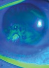
Technique of deep corneal foreign body removal
This article has been verified for CPD. Click the button below to answer a few short questions and download a form to be included in your CPD folder. The objective is to demonstrate a method for the safe and successful...

Like a moth to AC flare: CMV-associated hypertensive anterior uveitis
Hypertensive anterior uveitis can present a diagnostic challenge to clinicians working in emergency eye departments. While prompt initial control of intraocular pressure (IOP) and inflammation is essential, elucidating the underlying aetiology is critical for long-term visual outcomes. When there is...

Aqueous misdirection: a case series of unexpected surgical complications
Aqueous misdirection (AM), also known as malignant glaucoma, is a form of secondary glaucoma that typically presents with shallowing of the anterior chamber (AC), raised intraocular pressure (IOP), and reduced visual acuity (VA) in the presence of patent peripheral iridotomies...

Applanation tonometry in the pandemic era: Are facial masks an obstacle to a correct intraocular pressure measurement?
Current recommendations in the UK advise on wearing any facial covering to prevent the spread of the coronavirus [1]. Whilst this is vital for patient and hospital staff safety, it has led to several changes in the approach to a...
Recurrent corneal erosions secondary to isotretinoin use
Isotretinoin (13–cis-retinoic acid) is the first line treatment for moderate to severe nodulocystic or papulopustular acne [1,2]. Although it is a safe medication, it has several adverse side-effects, including ophthalmic manifestations, as shown in Table 1 [3]. These side-effects need...

Trabeculectomy with erroneous Mitomycin-C concentration – a near miss
Trabeculectomy is the most commonly performed surgical procedure for glaucoma in the United Kingdom and worldwide. Modifications to the technique have been made since its introduction in 1963, perhaps the most significant being the adjunctive use of mitomycin-C (MMC), which...

Fundus photography in Malawi – setting up a screening programmefor diabetic retinopathy
We present the case of a 53-year-old lady who presented to the diabetes outpatient clinic at Kamuzu Central Hospital (KCH), Lilongwe, Malawi. She was diagnosed with type 2 diabetes mellitus six years ago, for which she takes metformin orally. She...

Systemic sarcoidosis presenting with acute myopia and angle closure
Case report A 40-year-old Asian man presented to the Emergency Department with a one day history of sudden onset visual disturbance in his right eye. He complained of image distortion and noted that objects now appeared smaller. He also described...

Cyanoacrylate nail glue accidentally instilled into the eye instead of eye drops
Three hundred thousand cataract operations take place each year in the UK alone and each patient will receive a four week course of drops in order to cover them for any postoperative inflammation or infection. Accidental instillation of cyanoacrylate glue...

Bovine pericardium scleral patch graft associated scleritis: Ahmed valve implant for pupillary block glaucoma
Processed bovine pericardium is a lyophilised collagen sheet used as a surgical armamentarium. It renders the material antigenically inert with minimal inflammation [1]. Sclera patch pericardium graft (Tutopatch) is a collagenous membrane derived from solvent preserved, irradiated bovine pericardium [2]...

A refractive surprise after vitrectomy and capsulectomy
Figure 1: Right posterior capsule small aperture. We report a case of a pseudophakic patient who underwent vitrectomy and posterior capsulectomy. In spite of good visual acuity and absence of floaters, he was unhappy with the visual outcome. Case report...

Acute dellen formation post trauma
Corneal dellen are saucer-like thinnings, usually of the peripheral cornea [1]. Dellen formation is thought to be related to localised tear film instability [2], specifically the absence of the mucin component of the tear film. Without the mucin layer, dry...

