Introduction
Periorbital (sometimes called preseptal cellulitis) is a common condition which on its own is not normally an ophthalmic or surgical emergency, however it has the potential to cause severe and serious morbidity in cases where the infection has crossed the orbital septum into the orbit. This can occur if treatment is delayed and can lead to orbital cellulitis and risk of life-threatening complications like cerebral abscess, meningitis, cavernous sinus thrombosis and sepsis [1].
We report an interesting case of unexplained recurrent right-sided preseptal cellulitis in a relatively young female, which was found to be caused by a likely underlying herpes simplex virus eventually.

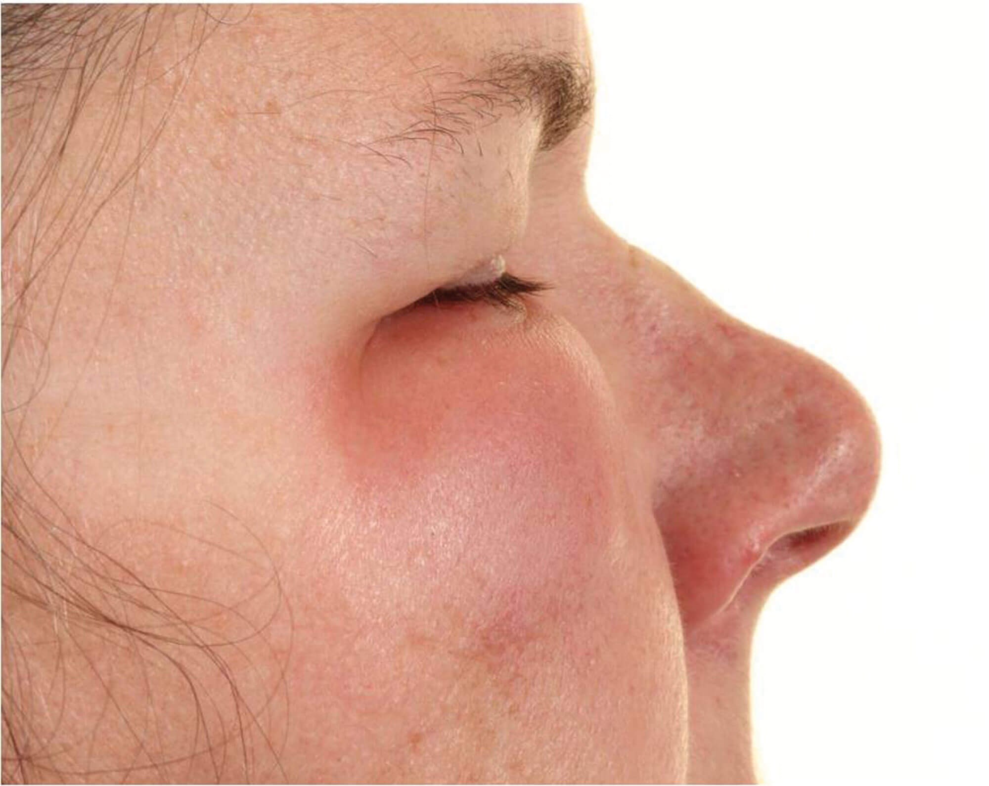
Figure 1: Right eye periorbital edema – lateral view.

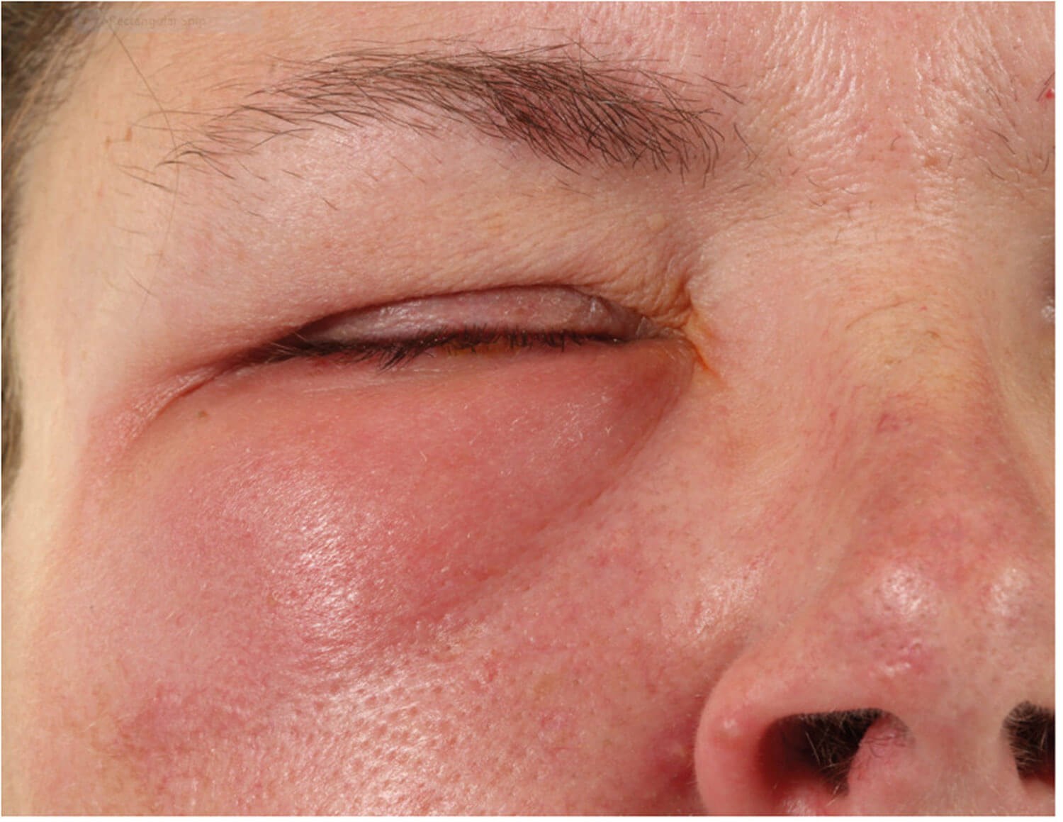
Figure 2: Right eye periorbital edema – anterior view.
Case
A female patient in her 40s was seen in the eye clinic at James Paget University Hospital (JPUH) in December 2019 with complaints of acute onset of right periocular swelling, watering and light sensitivity along with soreness and tenderness around the right eye for two to three days (Figure 1 and 2).
This was a recurrence of symptoms the patient had previously experienced on multiple occasions. She had an 18-year history of unexplained intermittent swelling around the right eye, each episode generally lasted for about two weeks and occurring approximately once every year. She was aware of a prodrome associated with these episodes starting with right lower lid swelling followed by a type of malaise, becoming run down and generally feeling low. Within a short period of time this would spread rapidly to the upper lid, becoming very swollen leading to closure of the eye. All her previous investigations in this regard including a CT scan of orbits were inconclusive. She mentioned that her previous episodes had subsided on occasions after taking antibiotics prescribed by different clinicians in the past.
On examination at the recent visit, her visual acuity was counting fingers on the right eye improving to 3/60 on pin hole measurement and 6/6 on the left eye. There was right periocular swelling with a tense, swollen and oedematous right lower lid, which was tender to touch (Figure 1 and 2). The conjunctiva on the right side exhibited papillae and follicles and there was a diffuse chemosis and some mild scleral inflammation. In the right cornea there was only punctate staining with fluorescein. Anterior chamber was quiet on examination with an intraocular pressure of 14mmHg. Fundoscopy and other eye examinations were normal.
Patient was investigated with complete blood picture, urea and electrolytes, a detailed immunology screen plus erythrocyte sedimentation rate (ESR) and C-reactive protein (CRP) These were all reported to be within normal range.
Treatment was commenced with oral coamoxiclav 625mg three times a day and a trial of oral steroids, prednisolone 30mg on a weekly tapering course which subsided the symptoms temporarily.

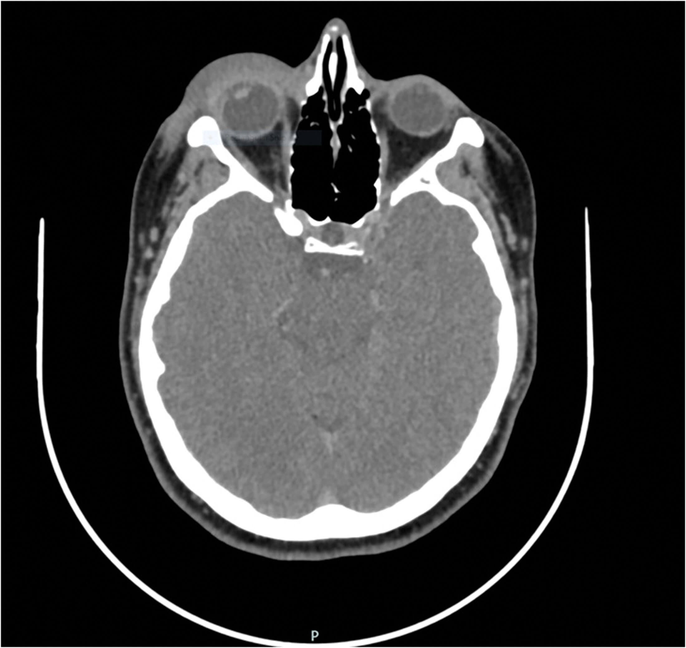
Figure 3: Right eye CT scan image showing periorbital soft tissue swelling mainly.
The case was discussed in the regional orbital multidisciplinary team (MDT) meeting with review of her orbital imaging. She had the initial CT scan of orbits and sinuses done in 2013 and a repeat scan with IV contrast in 2019. Both times the scan was done after most of her clinical signs had resolved, hence there was no significant pathology seen (Figure 3). The consensus of the MDT was the same, that there was no abnormality seen.
Differential diagnoses of preseptal cellulitis, inflammatory orbital disease and dacryoadenitis were considered, however, the normal blood tests and imaging ruled out dacryoadenitis and inflammatory orbital disease.
The patient had a recurrence in October 2020 with similar signs as before, however, a new muco-purulent discharge and right sided submandibular lympadenopathy were also noted at this visit, which prompted the clinician to carry out a right-sided conjunctival swab for viral, bacterial, and chlamydial culture and sensitivity. Microbiology was positive for herpes simplex virus (HSV) type-1. The patient was subsequently started on a high dose of oral antiviral treatment, valaciclovir 1gm three times a day for 10 days, followed by long-term low dose acyclovir prophylaxis which stabilised the symptoms and stopped the recurrences. The patient is kept on an extended interval follow-up in the local eye service and remains stable on the antiviral maintenance treatment.
Written consent was obtained from the patient to publish her case along with her images included in this report.
Discussion
Preseptal cellulitis is much more common in children and younger age groups, in contrast to orbital cellulitis, which more commonly affects adults [2].
The causative organisms for preseptal cellulitis are numerous, though staphylococcus aureus and streptococcus species are the most common pathogens [2].
Herpetic eye disease is usually caused by type 1 HSV which can manifest as blepharitis, canalicular obstruction, conjunctivitis, corneal complications, iritis, and retinitis in a varying degree and manner, however, HSV is commonly associated with herpetic keratitis, whilst herpes zoster virus (HZV) is associated with herpes zoster ophthalmicus more commonly [3,4].
HZV has been reported to be an uncommon cause of preseptal cellulitis [5].
The recommended treatment for preseptal cellulitis is oral antibiotics given empirically on the basis of assumption that the commonest causative pathogens are streptococcal and staphylococcal species. In our case, the patient was treated with oral antibiotics for her recurrent episodes, which led to a temporary improvement only.
HSV-associated ocular disease mostly affects the cornea and is usually treated with topical steroids and antiviral treatment like acyclovir [6].
In our case there was no significant corneal involvement, and the periocular tissues were predominantly affected, resulting in a unilateral preseptal cellulitis. Considering the viral etiology, the patient was treated with the standard antiviral treatment with a good clinical outcome.
In conclusion, we have presented an interesting case of recurrent unilateral preseptal cellulitis likely due to HSV‑1 virus with a several-year history of an unexplained cause. To the best of our knowledge, we believe this clinical scenario due to HSV infection has not been previously reported.
References
1. Bergin DJ, Wright JE. Orbital cellulitis. Br J Ophthalmol 1986;70(3):174-8.
2. Pandian DG, Babu RK, Chaitra A, et al. Nine years’ review on preseptal and orbital cellulitis and emergence of community – acquired methicillin-resistant Staphylococus aureus in a tertiary hospital in India. Indian J Ophthalmol 2011;59(6):431-5.
3. Kaye S, Choudhary A. Herpes simplex keratitis. Prog Retin Eye Res 2006;25(4):355-80.
4. Sanjay S, Huang P, Lavanya R. Herpes zoster ophthalmicus. Curr Treat Options Neurol 2011;13(1):79-91.
5. Qualickuz Zanan NH, Zahedi FD, Husain S. Varicella zoster causing preseptal cellulitis – uncommon but possible. Malays Fam Physician 2017;12(3):37-9.
6. Herpetic Eye Disease Study Group. A controlled trial of oral acyclovir for the prevention of stromal keratitis or iritis in patients with herpes simplex virus epithelial keratitis. Arch Ophthalmol 1997;115(6):703-12.
COMMENTS ARE WELCOME


