Case Reports archive for 2021
Usefulness of gonioscopy to investigate cause of corneal oedema after cataract surgery
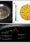
A 72-year-old man with ocular hypertension presented three months after routine right phacoemulsification and toric intraocular lens (IOL) implantation with a two-week history of an irritated right eye and a sudden deterioration in right vision. His preoperative spherical equivalence was...
Global health and conflict: the unseen consequences
Global eye health inequalities stem from poor access to affordable care, causing preventative vision impairment and blindness. In 2020, a study showed that 510 million people, the majority being in low-income and middle-income countries, had uncorrected near vision impairment simply...
A rare neonatal presentation of bilateral dacryocele and choanal atresia
Following a routine pregnancy, a newly delivered baby boy, born at term, was found to have increased work of breathing, stridor and a left medial canthal swelling. The baby required 100% oxygen via a face mask to maintain oxygen saturations....
An unusual presentation of sarcoidosis
*Equally contributing co-first authors. Case report A 45-year-old man presented to his local optometrist with a three-week history of severe intermittent left eye pain with associated blurred vision and tenderness around his left temple. Two days prior, he developed weakness...
Diabetes macular oedema in pregnancy self-resolving postpartum
*Equally contributing co-first authors. Diabetic macular oedema (DMO) is a common clinical presentation to ophthalmology clinics. Ample evidence exists for management of DMO in non-pregnant patients. However, there is a paucity of evidence on the optimal management of DMO in...
Macular imagery: observing the visual sensations pre- and post-Jetrea injections
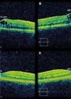
A 63-year-old woman, a professional painter, was diagnosed with vitreomacular traction (VMT) in 2017. She had a history of metamorphopsia, drop in visual acuity (VA) in the left eye (6/6 in the RE; 6/18 in the LE), foveal vitreomacular traction...
Iris chafing from displaced single-piece acrylic IOL
A 74-year-old man had persistent 3+ cell one month following left eye cataract extraction, complicated by anterior capsular rent and zonular dialysis at 7 o’clock, with single-piece acrylic intraocular lens implantation (IOL) in the capsular bag. Figure 1: Haptic-like transillumination...
Microcatheter in the vertebral artery as a cause of branched retinal artery occlusion?
A 19-year-old male presented to eye casualty with a seven-day history of a ‘blurred patch’ in the left eye. The patient denied any other visual symptoms including flashes or floaters and there had been no change in visual symptoms in...
Refractive surprise after cataract surgery caused by posterior capsular striae
Cataract removal with intraocular lens (IOL) implantation is one of the most frequently performed surgeries in current clinical practice [1,2]. New microsurgical techniques and refined IOL power calculations allow excellent refractive outcomes. Refractive surprise following cataract surgery is uncommon [1-3]...
Acute uveitis from late migration of soft lens matter 10 years post cataract surgery
A 58-year-old Caucasian male presented to the emergency eye clinic with a two-day history of a painful, red left eye and blurred vision. His past ocular history included uncomplicated left phacoemulsification cataract surgery in 2010 and left retinal detachment repair...
Lockdown and eye health – a case of accommodative spasm
A 25-year-old male presented to the eye casualty with a one-day history of sudden onset worsening vision. More specifically, he noted his vision was more blurred than usual and this was more exaggerated for near-work than for distance-work. He was...
Traumatic ‘toy’ gun injury leading to permanent vision loss
Pseudoxanthoma elasticum (PXE) is a progressive, inherited disorder of connective tissue that affects the skin, cardiovascular system and retina. Ocular manifestations of the disease are related to Bruch’s membrane, a thin elastic tissue layer located between the retinal pigment epithelium...


