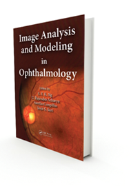
Recent advances have revolutionised ophthalmic imaging and helped understand the pathophysiology of ocular diseases and thus help in the diagnosis and management of ocular diseases. The authors of this book have gone through most of the available imaging techniques available for the eyes from the front to the back, including thermography, except ultrasound and CT and MRI scan.
The book acts as a bridging text between the actual physics and calculations used in imaging and the practical uses of these methods in understanding disease pathology. The calculations make the reading a bit dry to most ophthalmologists except those who are doing research or who are passionate about imaging.
Likening the eye to the conventional camera, Image Analysis and Modelling in Ophthalmology explores the application of advanced image processing in ocular imaging. This book does an outstanding job of describing established and new forms of retinal imaging, covering both acquisition and interpretation, information useful for clinicians as well as researchers at all levels interested in applications of retinal imaging, from students and residents to experienced retina specialists.
The chapters are well-written with relevant examples, and cover all the major parts of the eye including cornea, anterior segment, optic nerve and retina. Each chapter looks at the background of each one of these techniques, indications, advantages, disadvantages and limitations. It also assesses the use of these techniques in various diseases and its limitations. The authors discuss corneal topography, confocal microscopy and anterior segment imaging for anterior chamber diseases. Most of the chapters are devoted to the use of imaging in retinal diseases including fundus photography in diabetes, processing of images, optical coherence tomography, fundus fluorescein angiography, auto-fluorescence and vascular flow.
The authors introduce the A-Level set algorithm, explore the ARGALI system to calculate the cup-to-disc ratio (CDR), and describe the Singapore Eye Vessel Assessment (SIVA) system, a holistic tool which brings together various technologies from image processing and artificial intelligence to construct vascular models from retinal images. They discuss the advantages and disadvantages of different approaches to imaging the different structures.
There are a many textbooks written on ophthalmic imaging, but this book goes a bit further and describes the recent emerging techniques that are being used currently in only a limited number of centres. It gives you an insight into these new techniques. It helps in making an informed decision about use of these techniques in everyday practice.
I would recommend this book to clinicians and researchers who are involved in imaging but it may not be a good read for general ophthalmologists who may find this book quite daunting.


