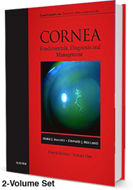
Originally published in 1997 as a comprehensive set of three volumes on cornea and external eye disease, this two volume edition, published at the end of 2016, offers “a complete multimedia resource, in print, online, eBook and video format”. There may be some practitioners who still refer on occasion to the original black and white edition, but this recent revised set has impressive high quality colour illustrations and well-presented text that is surprisingly compelling to read and content reflects current relevant topics.
As with earlier versions, the contributors are primarily based in the USA, but not exclusively as there are some contributions from Canada, South America, Europe, South East Asia, New Zealand and Australia.
Each volume is subdivided into parts, with the edges of the pages colour-coded for ease of navigation. Each of the 14 parts is subdivided into between one and nine sections. Each section comprises up to 15 chapters. A key concepts / points highlighted box introduces the chapters and there are numerous references accompanying each chapter. With such extensive referencing, there is likely to be the occasional error, but this should not cause any undue frustration. The index in each volume is comprehensive, includes both volumes and also provides the page numbers for the figures, tables and key concepts.
These books are likely to have broad appeal to all ophthalmic practitioners. The text deals with maintaining corneal integrity to facilitate optimal visual performance, managing the corneal tissue response to disease, or degeneration and trauma. There are also chapters concerned with the reconstruction of corneal tissue to preserve and enhance vision and minimise the potential social stigma that could accompany disfigurement from anterior segment trauma or pathology.
Volume one covers basic anatomy, physiology and pathophysiologic responses for the cornea, sclera and ocular adnexa. The other parts include examining and imaging techniques, differential diagnosis, conjunctival abnormalities, eye banking, diseases of the cornea including corneal trauma from biological and chemical warfare, applications of contact lens in corneal disease and diseases of the sclera and anterior uvea.
Volume two includes keratoplasty, therapeutic procedures, corneal collagen cross-linking, ocular surface transplantation and refractive surgery. The sections and chapters on newer techniques and procedures include anterior lamellar keratoplasty, endothelial keratoplasty, phakic intraocular lenses, with expanded sections on keratoprostheses, and ocular surface transplantation. In addition, some three hours of video clips supplement much of the content in the second volume including many of the more recent surgical techniques for example, deep anterior lamellar keratoplasty (DALK) and Descemet’s stripping automated endothelial keratoplasty (DSAEK).
Whether the reader is involved in the presurgical evaluation, the surgery, the postoperative management, or merely curious about all aspects that fall within the corneal specialty, there is undoubtedly a great deal of excellent material brought together under this title and time is well-spent delving into this highly recommendable update.
Inevitably, in order to include the most recent advances in knowledge of the cornea and associated ocular adnexa, some of the text referring to more obscure conditions found in the original publication has been excluded in this version. The specific part dealing with eye banking in this edition has been significantly condensed. This latest issue reflects the current developments in laser vision correction, replacing the role of radial keratotomy covered in earlier editions, but retains a chapter on incisional keratotomy in cataract surgery. However, these changes should not deter those considering purchasing these exceptional, high quality products that are excellent value for money.
As this text is unlikely to be updated for at least the next six years, based on previous timescales, it should be considered essential reference material, particularly if no earlier editions are available within the departmental library. It is likely to be popular with corneal and contact lens clinicians but also of interest for those working in ophthalmic accident and emergency roles, ophthalmic paediatrics, other medical and surgical ophthalmic specialities and those in academic, teaching or research departments.
The eBook version can be activated at no additional charge. Browsing the contents and viewing the enhanced images, both online and offline, is likely to be one of its attractions, however, access “is limited to the first individual that redeems the PIN on the cover of the book and may not be transferred to another party”. This may prove a disappointment if it is intended to be a shared resource within a department. Despite this, and accepting that the printed version comes as two large, heavyweight books, I have no hesitation in giving it a thoroughly deserved top rating.


