Pathology Quiz - Section Editor until Aug/Sep 2016
Latest Contribution
Jun/Jul 2015 Quiz
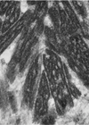
History A 35-year-old male presents with bilateral corneal opacities. Examination shows a honeycomb-type dystrophy. A penetrating keratoplasty is performed and the specimen sent for ophthalmic histopathological assessment. Figure 1 is the haemotoxylin & eosin (H&E). Figure 2 is a Masson...

Apr/May 2015 Quiz
History A 60-year-old man presented to the ophthalmologists with painful blurred left vision. Examination revealed a white vascular mass, occupying 60% of the anterior chamber space, arising from the iris. After conservative therapy, no useful vision remained. The eye was...

Feb/Mar 2015 Quiz 2
History A 60-year-old white Caucasian male, with a history of acne, presented with slate grey pigmentation of his upper forehead, pre-auricular skin, peri-oral area, forearms and shins. The conjunctivae showed bilateral lower tarsal conjunctival multiple black dots. One of these...

