This site uses cookies. By continuing to browse the site you are agreeing to our use of cookies.  Read our Cookies Policy.
Close
Eye News
  • Features
    • Close
    • Features
    • Allied Professions
    • Humanitarian
    • Interviews
    • AI & Oculomics
    • Ophthalmology
    • Optometry
    • Podcast videos
    • Supplements
  • Education
    • Close
    • Education
    • Learning Curve
    • Quiz
    • Top Tips
    • Trainees
    • Medico-Legal
    • The Truth Behind The Headlines
    • Case Reports
    • Pete's Bogus Journey
  • Reviews
    • Close
    • Reviews
    • Book Reviews
    • Journal Reviews
    • What's trending?
    • Tech Reviews
    • My Top Five
    • The Culture Section
  • Events
  • News
  • Product Guide
  • Industry News
  • Contact us
    • Close
    • Contact us
    • Write for Eye News
  • Home
  • Education
  • Quiz
  • Quiz 2 Feb/Mar 2015

Feb/Mar 2015 Quiz 2
By Hardeep Singh Mudhar

1 February 2015 | Hardeep Singh Mudhar | EYE - Cataract, EYE - Cornea, EYE - Glaucoma, EYE - Imaging, EYE - Neuro-ophthalmology, EYE - Oculoplastic, EYE - Oncology, EYE - Orbit, EYE - Paediatrics, EYE - Pathology, EYE - Refractive, EYE - Strabismus, EYE - Vitreo-Retinal, EYE - General
Share This


History


A 60-year-old white Caucasian male, with a history of acne, presented with slate grey pigmentation of his upper forehead, pre-auricular skin, peri-oral area, forearms and shins. The conjunctivae showed bilateral lower tarsal conjunctival multiple black dots. One of these dots was sampled (see Figure 1).
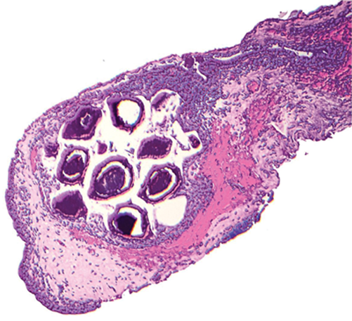


Figure 1.


Questions


  1. What type of epithelium lines the conjunctiva?
  2. What is seen in the histology image?
  3. Given the appearance in the figure, which questions
    should the patient be asked?
  4. What is the diagnosis?


Answers


1. Non-keratinising, goblet cell containing stratified squamous epithelium.
2. This shows black / brown concretions
in cysts.
3. Drug history!
4. These are minocycline pigmented concretions. This drug is well recognised to cause pigmentation of sun-exposed skin (increased melanin production) and cause pigmented concretions (iron and drug metabolites, not melanin). Many drugs are associated with conjunctival ‘pigmentation’ of which another example is chlorpromazine.


Further reading

1. Brothers DM, Hidayat AA. Conjunctival pigmentation associated with tetracycline medication. Ophthalmology 1981;88(12):1212-5.
2. Messmer E, Font RL, Sheldon G, Murphy D. Pigmented conjunctival cysts following tetracycline/minocycline therapy. Histochemical and electron microscopic observations. Ophthalmology 1983;90(12):1462-8.


COMMENTS ARE WELCOME

Share This
CONTRIBUTOR
Hardeep Singh Mudhar

HSOPS, Histopathology, Royal Hallamshire Hospital, Sheffield, UK

View Full Profile
Originally Published
EYE NEWS VOLUME 21 ISSUE 5 FEBRUARY/MARCH 2015
Download the full article
Download Article

Top Of Page

9 Gayfield Square, 
Edinburgh EH1 3NT, UK.

Call: +44 (0)131 557 4184
www.pinpoint-scotland.com

WEBSITE DETAILS
  • Cookie Policy
  • Data Protection Notice
  • Privacy Policy
  • Terms and Conditions
ABOUT US
  • Who we are
  • Register
  • Contact us
  • Contributors
  • Company Awards
DIGITAL ISSUES/GUIDELINES
  • Digital issues - Library
  • Supplements - Library
  • Guidelines
Accreditations
IPSO_FLAG_TEAL 2025.png cpdcertified.png

Pinpoint Scotland Ltd (Registered in Scotland No. SC068684) | © 2025 - Website by Gecko Agency