History
-
A 60-year-old man presented to the ophthalmologists with painful blurred left vision. Examination revealed a white vascular mass, occupying 60% of the anterior chamber space, arising from the iris. After conservative therapy, no useful vision remained. The eye was still painful and blind and enucleation was performed.
-
Figure 1 – sclera and choroid
-
Figure 2 – anterior chamber contents
-
Figure 3 – highest power of pathology in anterior chamber
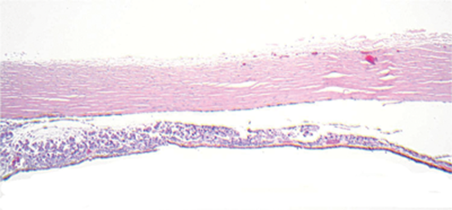
Figure 1.
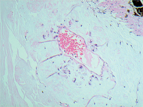
Figure 2.
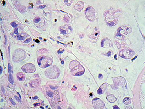
Figure 3.
Questions
1. What does Figure 1 show?
2. What is the bluish material filling the anterior chamber (Figure 2)?
3. What is shown in Figure 3?
4. How are Figures 2 and 3 related?
5. What is the diagnosis?
6. Which further investigations should be carried out?
Answers
1. This shows an abnormal infiltrate of cells in the choroid. Normal choroid is seen on the far right.
2. This is mucin.
3. These are atypical signet ring cells.
4. The signet ring cells contain intracytoplasmic mucin as well.
5. This is metastatic signet ring cell adenocarcinoma to the iris, anterior chamber and choroid.
6. This patient required detailed body imaging to search for a primary lesion.
Discussion
Metastases are the commonest intraocular tumour. Ninety percent present in the choroid, 10% iris and ciliary body because the uvea is a vascular bed. In the paper by Shields et al. examining 520 cases (Ophthalmology 1997;104:1265-76) breast was the commonest primary site – 47%, followed by lung 21%, gastrointestinal tract (GIT) 4%, skin melanoma 3%, kidney 2%, prostate 2%, others 21%.
Twenty-five percent of cases of patients with uveal metastases have no history of primary cancer – the ophthalmologist is the first to make a diagnosis of metastatic disease, as in this case.
Outcome of this case: imaging showed a signet ring cell primary lung adenocarcinoma.
COMMENTS ARE WELCOME


