History
-
A 35-year-old male presents with bilateral corneal opacities. Examination shows a honeycomb-type dystrophy. A penetrating keratoplasty is performed and the specimen sent for ophthalmic histopathological assessment.
-
Figure 1 is the haemotoxylin & eosin (H&E).
-
Figure 2 is a Masson trichrome stain.
-
Figure 3 is a transmission electron micrograph of the pathology shown in Figures 1 and 2.
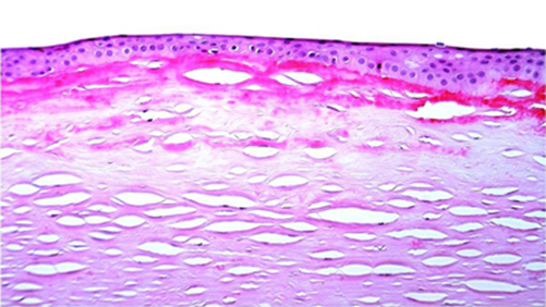
Figure 1.
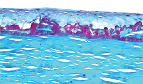
Figure 2.
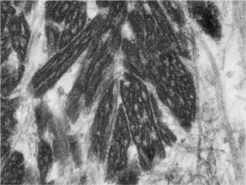
Figure 3.
Questions
1. What are the key features on the H&E?
2. What does the Masson trichrome show?
3. What does the electron micrograph display?
4. What is the likeliest diagnosis?
5. What is the H&E differential diagnosis?
Answers
1. Sub-epithelial eosinophilic diffuse deposit with some in the anterior stroma, with epithelial atrophy. 2. Masson’s trichrome positive deposits matching the distribution on the H&E.
3. Electron dense rhomboidal deposits.
4. This is a good example of Reiss Buckler dystrophy (BIG-H3 family), which is essentially an anterior variant of granular dystrophy.
5. The main H&E differential is Thiel Behnke dystrophy, secondary amyloid deposits and primary gelatinous drop-like dystrophy. Thiel Behnke shows thin curly fibres on electron microscopy; amyloid deposits are Congo Red positive and show a distinctive fibrillar architecture on electron microscopy.
COMMENTS ARE WELCOME


