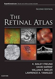
Those readers familiar with the first edition of The Retinal Atlas will appreciate the comprehensive nature of this publication, and this latest version incorporates retinal images generated by recently developed imaging technology.
As the title suggests, this hardcover version is not a pocket guide, however, as a bonus, one can activate the eBook version of this title at no additional charge for use on a desktop or a mobile device. Browsing the contents and viewing the enhanced images, both online and offline, is one of its attractions. The diversity of conditions covered is stunning, each one concisely described and accompanied by high quality illustrations.
There are 15 chapters plus an index. The opening chapter briefly describes the normal retinal histology, the fundus appearance with colour photography, fluorescein angiography (FA) indocyanine green (ICG) fundus autofluorescence (FAF) ultra widefield imaging, optical coherence tomography (OCT), and optical coherence tomography angiography (OCTA). The next chapter covering hereditary chorioretinal dystrophies is the longest. Each chapter, except the first, is supported by an extensive suggested reading list, which is particularly useful as the text describing each condition (many of which may be rarely encountered) is limited.
The chapters include Paediatrics, Inflammation, Infection, Retinal Vascular Disease, Degeneration, Oncology, Vitreomacular Traction, Epiretinal Membranes and Macular Holes, Traumatic Chorioretinopathy, Complications of Ocular Surgery, Chorioretinal Toxicities and Congenital and Developmental Anomalies of the Optic Nerve. This latter chapter has a comprehensive reading list.
The chapter pages are colour-coded along the edge for ease of navigation. The profusion of illustrations, generated from several different modalities of imaging technologies are colour and shape-coded to aid interpretation. OCT images, for example, have a broad black border in a square frame.
This book is a pleasure to browse, visually attractive, easy to navigate and an encyclopaedia of retinal disease, although in the hardback version, it’s quite a hefty tome to hold. The only, very minor, criticism one might consider is the lack of any suggested reading list for the first chapter that could direct the naïve reader to publications which describe the new imaging technology and how to interpret the images they generate. Despite this, it is likely to be popular with any student of retinal diseases, from the least to the most experienced practitioner.


