This site uses cookies. By continuing to browse the site you are agreeing to our use of cookies.  Read our Cookies Policy.
Close
Eye News
  • Features
    • Close
    • Features
    • Allied Professions
    • Humanitarian
    • Interviews
    • AI & Oculomics
    • Ophthalmology
    • Optometry
    • Podcast videos
    • Supplements
  • Education
    • Close
    • Education
    • Learning Curve
    • Quiz
    • Top Tips
    • Trainees
    • Medico-Legal
    • The Truth Behind The Headlines
    • Case Reports
    • Pete's Bogus Journey
  • Reviews
    • Close
    • Reviews
    • Book Reviews
    • Journal Reviews
    • What's trending?
    • Tech Reviews
    • My Top Five
    • The Culture Section
  • Events
  • News
  • Product Guide
  • Industry News
  • Contact us
    • Close
    • Contact us
    • Write for Eye News
  • Home
  • Reviews
  • Book Reviews
  • Multimodal Retinal Imaging

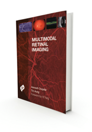
Multimodal Retinal Imaging
Reviewed by Jaya Myneni

1 December 2015 | Jaya Myneni | EYE - Imaging, EYE - Vitreo-Retinal

Understanding of rapidly advancing retinal imaging techniques is important as they have changed the management of retinal conditions considerably. Interpretation of these tests is a vital skill in the armamentarium of every practising ophthalmologist.

The book is directed at general ophthalmologists and doctors in training for a good, up-to-date overview of the subject. It is well illustrated and written in a consistent format by multiple authors.

The book is organised into two main parts. In the first part, imaging techniques like fluorescein and indocyanine green angiography, optical coherence tomography, ocular echography, autofluorescence imaging and optic nerve head imaging are discussed in seven chapters. Principles and techniques of angiography and optical coherence tomography are outlined.

The chapter on ocular echography not only describes the principles but also details the techniques of examination, which is particularly useful for trainee ophthalmologists and allied professionals. Scanning laser ophthalmoscopy and clinical applications have been summarised. Setting up of an imaging centre, policies, operational issues and staff management are also discussed.

The second part focuses on assessment of some common conditions like age-related macular degeneration, vascular disorders, diabetic retinopathy and some uncommon conditions like dystrophies and neoplasms. The chapters succinctly cover pathophysiology, clinical presentation and findings on various modalities of angiography and imaging. Imaging techniques for disc assessment in glaucoma are compared and presented in a separate chapter. Many common vitreoretinal conditions which were omitted from the book could have been included to make it more comprehensive.

The book is concise with an attractive layout and good quality clinical pictures, making it easy to read and useful for trainee ophthalmologists.

Share This
CONTRIBUTOR
Jaya Myneni

Royal Liverpool University Hospitals, UK.

View Full Profile

Book Details

Reviewed by: Ms Jaya Myneni, Specialty Trainee in Ophthalmology, Royal Liverpool University Hospitals, UK.


Editor: Amresh Chopdar, Tin Aung


Published by: JP Medical


Format: Hardback


Extent: 169 pages


ISBN: 978-1-907816-60-4


Price: £90.00


Value Rating: 3/5


BUY THE BOOK
Archive
  • 2025
  • 2024
  • 2023
  • 2022
  • 2021
  • 2020
  • 2019
  • 2018
  • 2017
  • 2016
  • 2015
  • 2014
  • 2013

Top Of Page

9 Gayfield Square, 
Edinburgh EH1 3NT, UK.

Call: +44 (0)131 557 4184
www.pinpoint-scotland.com

WEBSITE DETAILS
  • Cookie Policy
  • Data Protection Notice
  • Privacy Policy
  • Terms and Conditions
ABOUT US
  • Who we are
  • Register
  • Contact us
  • Contributors
  • Company Awards
DIGITAL ISSUES/GUIDELINES
  • Digital issues - Library
  • Supplements - Library
  • Guidelines
Accreditations
IPSO_FLAG_TEAL 2025.png cpdcertified.png

Pinpoint Scotland Ltd (Registered in Scotland No. SC068684) | © 2025 - Website by Gecko Agency