
Ophthalmology

25 years of OCT
David Huang first described optical coherence tomography (OCT) in 1991, in his seminal paper on the subject in Science. This method developed the work of others on ophthalmic interferometry, which essentially showed that measuring reflected light could be used to...

High quality retinal image grading and management service by the NetwORC UK
In 2004 a network of three ophthalmic reading centres in Belfast, London and Liverpool (known as NetwORC UK) was established to form the largest reading centre in Europe for the purpose of providing high quality grading of ophthalmic images for...

Optical coherence tomography – reinventing the eye examination
It has been 25 years since Huang et al. presented the first optical coherence tomography (OCT) images in Science [1]. With vast improvements in OCT technology over the years, it is now possible to acquire high-resolution cross-sectional images of the...

The refractive index in the eye lens – implications for clinical practice and optical design
The eye may appear to be a comparatively simple organ and yet its optical system is complex and continues to be a source of investigation and research. The major optical elements are considered to be the cornea and the lens...
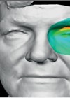
Can 3D facial imaging improve patient management in disfiguring eye disease?
Fight for Sight is the leading UK charity funder of eye research. Since the 2013 Sight Loss and Vision Priority Setting Partnership we have encouraged our researchers to work on priorities jointly identified by people affected by sight loss and...

Open source and tele-manufacturing for ophthalmology
Open source or crowd-sourcing and crowd-collaboration are concepts almost always associated with software and public online projects such as Wiki project. Never had I imagined that my team would apply the same principle in ophthalmology. Just less than a month...

Anterior segment imaging: a photographer’s view
My name is Rosalyn Painter and I work within the vision science and ophthalmic imaging team at Bristol Eye Hospital, where we cover all aspects of imaging within the hospital, including fluorescein angiograms, fundus photography, optical coherence tomography (OCT), slit-lamp...

What’s next in retinal imaging? Faster, deeper and full-on
Fast-evolving technological leaps are opening the way toward clinically useful ocular coherence angiography, generating 3-dimensional microvasculature maps without intravenous dye injection, as well as whole-eye imaging, handheld patient-operated optical coherence tomography (OCT) devices and, for challenging vitreoretinal procedures, integrated intraoperative...

