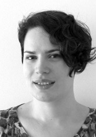
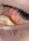
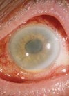
My name is Rosalyn Painter and I work within the vision science and ophthalmic imaging team at Bristol Eye Hospital, where we cover all aspects of imaging within the hospital, including fluorescein angiograms, fundus photography, optical coherence tomography (OCT), slit-lamp photography and many more techniques.
My specialist interest lies in the field of anterior segment imaging including slit-lamp photography. I work closely with three consultants and have a good working relationship with them and the rest of the anterior segment team, this enables me to gain a thorough understanding of what each consultant needs from the imaging they request and the subtle differences in why they want things done in a specific way. When I finished university I was lucky to promptly find a position in a high street optometrist and soon progressed to imaging for refractive laser eye surgery. I was very fortunate to work with a surgeon who liked to teach and would often call me into her clinic room to show me interesting conditions on the slit-lamp.
This experience boosted my confidence and motivated me to apply for a position at Southampton Eye Unit where I received most of the technical training for the position I now hold. Nearly all the training we receive is on the job; teaching from peers as well as lots of practice and research of my own. When we receive new equipment there is always industry training, the specialist industry trainers are always happy to help with queries once the equipment is installed. The Ophthalmic Imaging Association (OIA), of which I am on the committee, has teaching days several times a year, which are very useful for learning new techniques and sharing ideas.
For my own continuous professional development I have since presented at conferences and written a paper on slit-lamp photography for the Journal of Visual Communications in Medicine. This experience has given me a passion for anterior segment imaging which I hope to share with you.
I am able to follow my passion because I work with a team of highly skilled professionals who are equally passionate about their work. We teach each other new skills and have open discussion about techniques. I know that if I did not work with these people I would not have the scope for personal improvement that I do. Within the team we have specialists in medical retina, research imaging and glaucoma, as well as other members of the team who are looking to discover which element of ophthalmic imaging is their niche. The team are capable of operating all imaging equipment so that the service can be run when unforeseen absences occur (or even just to cover a day’s holiday) without affecting the patient experience.
Being able to specialise doesn’t mean that is all we do, it just means we are more closely involved with different parts of the service.
On a day-to-day basis I work in a room with an anterior segment slit-lamp camera, an anterior segment OCT, a specular microscope and a Pentacam tomographer. This equipment allows me to image the structure of the anterior segment in great detail. I also have experience with using a confocal microscope. Bristol Eye Hospital is a leading centre for the diagnosis and treatment of Acanthamoeba, as well as fungal keratitis, and a confocal microscope is essential for early diagnosis in these conditions.
The equipment that gives me the most satisfaction is undoubtedly the slit-lamp camera. I have the opportunity to show a patient’s condition and track it over time, as well as photographing what can be a traumatising condition for the patient in a way that can help them understand what is happening and come to terms with it. Imaging is important for patients to feel comfortable with what is happening to them as it is often not obvious because subtle changes can have sight-threatening impacts. It is my job to help document this. I often get patients exclaim with wonder when they see their images; this makes my job enjoyable and rewarding. Of course you get the patients that don’t want to know what is happening or what the condition looks like and I understand and respect that. These patients understand why we do the imaging; they just don’t need to see it for themselves.
As I am able to work closely with a small group of consultants I can aim to deliver a service that provides them with the tools they need to diagnose and monitor conditions and they have someone in a position to know what is needed even when the clinic is pressured for a detailed explanation. I often see the patient before the doctor; the imaging is done in this way to improve the flow of the clinic. I therefore have to understand the conditions and how to image them fully so the patient doesn’t have to come and see me again after they have seen the doctor.
Some of the specialist clinics I work with are the Boston keratoprothesis service, the Acanthamoeba keratitis services and the corneal collagen crosslinking and refractive surgery service with Consultant Ophthalmic Surgeons Mr Derek Tole, Mr Stuart Cook and Mr Phil Jaycock. I will be involved with the imaging for up and coming corneal graft research projects as well. Very occasionally we are called to theatre to image a procedure that is particularly interesting so it is advantageous to have a thorough understanding of a condition before entering the operating theatre, this is one area specialising can make a difference as you know what to expect, where you can stand to not be in the way and when is an appropriate time in the procedure to take a photograph. You don’t want to be distracting the surgeon with a flash or a question at an inopportune moment.
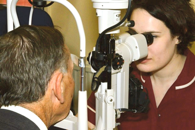
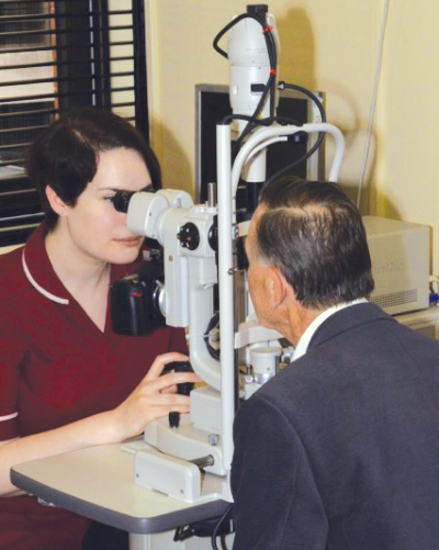
Slit-lamp camera examination.
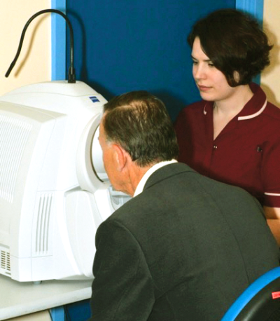
Anterior segment Visante OCT.
As we are a teaching hospital, images that are taken in clinic are often used for presentations and teaching, patients are informed and consent is gained for images to be used. Some of our doctors rotate around specialities on a regular basis therefore it is helpful for some consistency of imaging and nursing staff to help this process. If we can provide a service where a patient does not realise that it is a doctor’s first or last day in a rotation we are doing something right. We are there to support the medical team with a multi-disciplinary approach, as well as providing patient care.
There can be consistency for the patient and a rapport is built up over time with the patients that become regular attenders. I find this helps me get better images, as the patient is relaxed. When I’m not in clinic, and the service is being provided by a colleague, the patient still gets the same level of care. It is the same when I work in the retinal clinic, I may not know the patient as well as my colleague but I aim to give the same level of care and professionalism as they would expect from someone they have seen before.
We are also fortunate enough to be trained to see patients in a virtual clinic setting at peripheral sites. We perform exams and measurements that are then fed back to optometrists at Bristol Eye Hospital who review the notes and images and then make decisions based on these findings.
The patients we see in these clinics are routine follow-ups who do not need regular attention by a doctor or shared care optometrist but who do need review once a year or so.
To be able to give the quality of care we expect when we ourselves visit an outpatient setting we must all work closely as a team. The imaging and nursing teams here aim to communicate and work together to provide patients with consistency and care, this is not always easy in such a busy environment so knowing the team you are working with is vital. I know when I need to help with duties that are not necessarily part of my role it helps the clinics run smoothly and supports the rest of the team.
My role has developed over the last year through my own research and advice from clinicians, as well as expanding clinics allowing for more exposure to different conditions. The role, as with all imaging positions, is based on continuous learning and development with the service. Before I started in my role the anterior segment imaging was staffed by request from the clinician, a member of the imaging team had to come to the clinic, where the equipment is based, every time imaging was required. Now that I am permanently based in the clinic, requests come directly to me, this has allowed for clinics to be more efficient as there are not two flights of stairs between the imager and the patient and equipment but also allowed for more anterior segment imaging to take place as there is a dedicated imager in clinic.
Advances in technology have made my job more interesting and challenging. I find that the more access to a service, the more demand there is for it. Unfortunately funding is not always available to update every bit of equipment every time there is an advance in technology, this is not economically viable so we work with what we have and learn to get the best out of it.
In a growing service I feel that specialisation is necessary and a very welcome development in my role. I can learn and teach and develop a service I can be proud of, this isn’t possible without the support of all the staff within the hospital. I could not do my job effectively without the help of the medical secretaries, booking coordinators, medical records staff and cleaners – staff members that are not necessarily seen by the public but the hospital would be lost without them.
Working in a team as large as we have at Bristol Eye hospital I am fortunate enough to be given the opportunity to specialise in anterior segment imaging. I have a passion for this and I know how lucky I am to be in this position.
TAKE HOME MESSAGE
-
I have the opportunity to show a patient’s condition and track it over time. I can photograph what can be a traumatising condition for the patient in a way that can help them understand what is happening and how to come to terms with it.
-
I often get patients exclaim with wonder when they see their images; this makes my job enjoyable and rewarding.
-
Some of our doctors rotate around specialities on a regular basis, therefore, it is helpful for some consistency of imaging and nursing staff to help this process. If we can provide a service where a patient does not realise that it is a doctor’s first or last day in a rotation we are doing something right!
Declaration of Competing Interests: None declared.
COMMENTS ARE WELCOME


