This article has been verified for CPD. Click the button below to answer a few
short questions and download a form to be included in your CPD folder.

The objective is to demonstrate a method for the safe and successful removal of a metallic foreign body (FB) located at the level of Descemet’s membrane. While superficial metallic corneal FBs and intraocular FBs are common in ophthalmic emergency settings, an FB positioned just above Descemet’s membrane is a notably rare presentation.
Case report
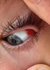
A male in his 30s, who was asymptomatic, presented to our eye casualty after noticing a black spot on his right cornea, most likely related to a welding incident a couple of weeks prior. The visual acuity was normal and slit lamp examination confirmed a deep stromal metallic FB situated just above Descemet’s membrane (Figure 1). He had a past ocular history of a few superficial FB removals in outpatient settings, with negligible residual corneal haze in paracentral and peripheral locations. The main cause of these incidents was not wearing protective goggles.

Figure 1: OCT showing preoperative deep corneal FB.
The patient was booked to be seen by a corneal consultant in the central unit for further investigations the next morning. An anterior segment optical coherence tomography (OCT) and corneal tomography confirmed that the FB was located just above Descemet’s membrane (Figure 1). The rest of the examination was unremarkable, with no signs of infection, and the Seidel sign was negative. The prognosis was guarded due to the risk of scarring and astigmatism, which was explained to the patient during the preoperative consultation, and informed consent was obtained.
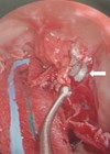
The local cornea team proceeded with the surgical removal of the FB in the operating theatre as a day-case under local anaesthesia two weeks later. The procedure involved creating a cross-shaped (‘X’) corneal incision to gain adequate access to the FB. A 30G needle was bent to ensure the FB could be safely and successfully removed via a lateral approach. A bandage contact lens was placed for protection and to aid in the healing process.
Discussion
Metallic corneal FBs are a common ophthalmic emergency. Corneal FBs typically involve the anterior stroma and are usually easily removable in an outpatient setting. However, deep stromal FB removal often requires surgical intervention.
Patients with corneal FBs commonly present to eye emergency departments or ophthalmic care centres, such as opticians [1-3]. The most frequent cause of corneal FBs is high-velocity mechanical work, such as hammering, grinding and drilling metals. These activities often result in FBs becoming lodged in the eye. Patients may present with acute symptoms, including pain, discharge, light sensitivity, blurred vision, epiphora, ptosis and conjunctival injection.
The cornea is the dome-shaped, windscreen-like covering of the eye, richly innervated and composed of clear tissue [4]. Its transparency is crucial for focusing light onto the retina, which is necessary for clear vision. The cornea provides almost 67% of the eye’s focusing power. Any corneal trauma may cause both physical and functional disturbances to the eye. Initially, corneal edema may cause photophobia and reduced visual acuity. Over time, corneal astigmatism resulting from scarring or irregularities may persist, especially when FBs are deeply embedded, leading to significant visual deterioration [5].
The cornea is a protective window for the eye [6]. The eyelid, tear film and bony orbital walls play vital roles in protecting the ocular surface from damage. The blinking mechanism of the eyelids and lashes produces reflex tears, which help reduce the impact of foreign debris on the cornea. Crucially, perforating or penetrating ocular injuries can lead to severe outcomes such as endophthalmitis or retinal detachment, potentially resulting in permanent blindness [7].
Corneal FBs can result from either blunt or penetrating trauma. Traumatic injuries to the anterior segment may involve lid abrasions, corneoscleral tears, corneal ulcers, iridodialysis, anterior uveitis, traumatic mydriasis, relative afferent pupillary defects, anterior or posterior capsular rupture, traumatic or rosette cataracts, lens dislocation, cortical matter disturbance, zonular dialysis, and vitreous prolapse. Posterior segment injuries may include vitreoretinal complications such as retinal tears, choroidal or retinal detachment, vitritis, and intermediate or panuveitis [8].
The incidence of corneal FBs varies in the literature. A Swedish study reported an eye injury rate of 8.1 per 1000 individuals, with 40% of these injuries involving corneal or conjunctival FB. In this study, data from a field hospital during the 1991 Gulf War showed that 14% of injuries involved ocular trauma, and among these, 17% involved corneal FBs. Notably, only 3% of the injured patients were wearing their issued protective goggles [9].
Several published studies have documented the management of deep corneal FBs, with surgical removal being the standard approach when FBs are embedded within the deeper stroma. A study by Sharma, et al. presented a case of a wooden FB embedded in the stroma, where the surgical procedure involved the creation of a corneal track to access the FB with the aid of a 26G needle. The FB was then trapped in the concavity of one limb of vitreoretinal FB forceps, a technique similar to that used in our case, though additional forceps were utilised. This approach was found to be effective with minimal complications and favourable outcomes in terms of visual acuity, no corneal ulcer formation, and preserved corneal integrity [10].
In another case series by Das D, et al., a group of patients with impacted corneal FBs over a 50-year period were classified based on the level of impaction, and various management options were discussed. The study highlighted that the most common cause of this condition is workplace-related injury, as seen in our case. They noted that management varies by patient and depends on several factors, which aligns with the surgical management provided to our patient and prompted us to innovate a new technique for removing the FB from an unusual location [11].
Similarly, a study by Duan, et al. emphasised the risk of significant corneal infection and keratopathy following the removal of deep FBs. They recommended careful postoperative submission of FBs for fungus and bacterial cultures, along with proper medical treatment if necessary. The study further confirmed the successful technique of creating a corneal tunnel with a keratome to reach the FB, followed by removal with a self-made device using a suture needle. We also performed a culture, which was essential in planning postoperative medical treatment, and fortunately, the culture report came back negative for infection, allowing us to stop prophylactic antibiotics [12].
The study by Donnenfeld, et al. suggested that a bandage soft contact lens may be considered to relieve pain, speed recovery, correct vision and reduce surface disruption associated with blinking following traumatic corneal abrasions. However, caution should be exercised if infection is a concern. As in our case, there was no infection, and the corneal surface disruption was prevented with a bandage soft contact lens for a speedy recovery. A protective shield was applied instead of a patch, resulting in a successful outcome [13].
These cases demonstrate a consistent approach to the management of deep corneal FBs, where surgical removal under local anaesthesia, followed by postoperative care with protective measures (including a bandage contact lens), plays a crucial role in minimising complications and improving visual outcomes.
Our case differs from many reported cases, as the metallic FB was located just above Descemet’s membrane, with the anterior cornea fully healed and smooth at the time of presentation. Typically, FBs in deeper stromal layers lead to visible scarring and irregularities of the anterior corneal surface. However, in our patient, the corneal surface had already healed, which increased the challenge of surgical removal. An X-shaped corneal incision was necessary to lift the FB. We also emphasised the risk of significant corneal scarring and astigmatism following the removal of deep FBs and provided careful preoperative counselling regarding potential visual outcomes.
Our case further highlighted the utility of advanced imaging techniques, such as anterior segment OCT, in assessing the depth and position of FBs, which was essential in planning the surgical approach. Despite the complexity of the surgical approach, favourable outcomes were achieved, suggesting that early detection and surgical intervention can result in positive results, even when the FB is embedded deep within the stroma.

Figure 2: Postoperative tomography map with 0.3D astigmatism.
Conclusion
The deep corneal metallic FB was successfully removed, as shown in the tomography map with 0.3D of astigmatism (Figure 2). No postoperative complications were observed during the weekly follow-up visit. The patient was advised to resume his routine activities and work with protective goggles a few days later. The ocular integrity, visual acuity and corneal shape were all maintained, as shown in the anterior segment OCT scan (Figure 3) taken five months after surgery.

Figure 3: OCT showing postoperative corneal scarring.
The removal of deep metallic FBs is challenging due to the high risk of Descemet’s membrane rupture and displacement into the anterior chamber.
Removing an FB through the anterior chamber should be avoided to prevent complications. This lateral approach technique is safe, easy to perform, and offers a good prognosis. Corneal specialist referral is necessary for better outcomes, and the procedure should be performed in an operating theatre.
References
1. Zimmerman DR, Shneor E, Millodot M, Gordon-Shaag A. Corneal and conjunctival injury seen in urgent care centres in Israel. Ophthalmic Physiol Opt 2019;39(1):46–52.
2. Sen E, Celik S, Inanc M, et al. Seasonal distribution of ocular conditions treated at the emergency room: a 1-year prospective study. Arq Bras Oftalmol 2018;81(2):116–9.
3. Samoilă O, Ostriceanu S, Samoilă L. Epidemiology of ocular emergencies in Cluj ophthalmology clinic. Rom J Ophthalmol 2016;60(3):165–9.
4. Yang AY, Chow J, Liu J. Corneal Innervation and Sensation: The Eye and Beyond. Yale J Biol Med 2018;91(1):13–21.
5. Feizi S. Corneal endothelial cell dysfunction: etiologies and management. Ther Adv Ophthalmol 2018;10:2515841418815802.
6. Eghrari AO, Riazuddin SA, Gottsch JD. Overview of the Cornea: Structure, Function, and Development. Prog Mol Biol Transl Sci 2015;134:7–23.
7. Turvey TA, Golden BA. Orbital anatomy for the surgeon. Oral Maxillofac Surg Clin North Am 2012;24(4):525–36.
8. Mohseni M, Blair K, Gurnani B, Bragg BN. Blunt Eye Trauma. Treasure Island, Florida; StatPearls Publishing; 2025.
9. Heier JS, Enzenauer RW, Wintermeyer SF, et al. Ocular injuries and diseases at a combat support hospital in support of Operations Desert Shield and Desert Storm. Arch Ophthalmol 1993;111(6):795–8.
10. Sharma A, Sharma R. Retrieval of deep corneal stromal wooden foreign body using vitreoretinal foreign body forceps as scoop: An innovative technique. Indian J Ophthalmol 2023;71(12):3718–20.
11. Das D, Agarwal S, Raj JS, et al. Management of impacted corneal foreign bodies: A review. IP Int J Ocul Oncol Oculoplasty 2021;7(2):131–8.
12. Duan H, Yan S. Observation of clinical efficacy after surgical removal of deep corneal plant foreign bodies. Eye Sci 2013;28(1):30–3.
13. Donnenfeld ED, Selkin BA, Perry HD, et al. Controlled evaluation of a bandage contact lens and a topical nonsteroidal anti-inflammatory drug in treating traumatic corneal abrasions. Ophthalmology 1995;102(6):979–84.
Declaration of competing interests: None declared.