This article has been verified for CPD. Click the button below to answer a few
short questions and download a form to be included in your CPD folder.

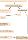
Neuromonitoring, for example in conditions causing raised intracranial pressure (ICP) such as traumatic brain injury (TBI), space-occupying lesions and idiopathic intracranial hypertension (IIH), remains challenging.
Current methods rely heavily on invasive monitoring using ICP probes, which carry risk of infection and haemorrhage [1,2]. While imaging such as CT and MRI can be helpful, they are timely and require transport of potentially unstable patients.
Ophthalmic examination, including pupillary responses, assessing the optic disc and spontaneous venous pulsations (SVP) can provide crucial information about ICP and cerebral perfusion. In fact, the retina and brain share a common embryological origin, from the neural ectoderm, resulting in similar vascular characteristics [3]. For example, both vascular beds demonstrate comparable autoregulation mechanisms, responding to arterial perfusion pressure and metabolites [3]. The retinal vasculature, which arises directly from the ophthalmic artery, is a branch of the internal carotid [3].
Considering this, imaging modalities, such as optical coherence tomography (OCT) and OCT angiography (OCTA) offer a promising non-invasive alternative that leverages the anatomical and physiological relationship between the retina and cerebral circulation. This article summarises methods in which the eye can be used as a proxy for cerebral assessments, focusing on established assessment methods and prospective tools.
Non-invasive ophthalmic assessments
Pupillary responses
Basic neuro-observations include examination of pupillary responses. Pupillary responses provide real-time information on brainstem function and the presence of space-occupying lesions or raised ICP. For example, studies have demonstrated that even in the absence of focal pathology, TBI can cause delayed, slowed or reduced pupillary light responses while maintaining symmetry [4]. Moreover, asymmetric pupils could suggest focal cerebral pathology. For instance, unilateral mydriasis and pupil non-reactivity classically indicate oculomotor nerve compression, often from herniation due to raised ICP or intracranial lesion affecting this cranial nerve and its nucleus [5]. Third nerve palsy is accompanied by ptosis and down and out eye positioning, and recognising that these ophthalmic signs could represent cerebral pathology is key in clinical assessment, although they may be less noticeable when patients are unconscious [5]. Furthermore, relative afferent pupillary defects (RAPD) indicate optic nerve dysfunction, such as from demyelinating disease, compression and ischaemia, further aiding cerebral assessment through bedside examination.
Papilloedema
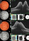
Papilloedema is oedema of the optic disc caused by raised ICP [6]. Elevated ICP transmitted along the subarachnoid space of the optic nerve causes stasis of axoplasmic flow and optic nerve fibre swelling [7]. Clinically, this manifests as symptoms of transient visual obscuration, blurred vision, pulsatile tinnitus, diplopia (from sixth nerve palsy) and headache, and can be diagnosed on fundoscopic examination and OCT [7].
However, there are several limitations to consider, including the fact that papilloedema is a delayed manifestation of high ICP. A prospective study by Steffan, et al. demonstrated that in patients with acute ICP elevation of 20–30mmHg from trauma or intracranial haemorrhage, papilloedema did not develop across assessments over three consecutive days [8]. Moreover, in one study of acute TBI, only one in seven patients showed disc swelling at pressures of 30–70mmHg, demonstrating that the absence of papilloedema does not exclude ICP elevation, particularly in the acute setting [8].
In addition, pseudo-papilloedema is a key differential diagnosis, where the optic disc may appear swollen despite the absence of nerve fibre layer oedema, for instance in cases of optic disc drusen and congenital disc anomalies [9]. Fundus autofluorescence, assessed using fundus photography or OCT laser devices, can reveal superficial drusen; cross-sectional OCT imaging of the optic nerve head can detect superficial buried drusen; and B-scan ultrasonography can aid detect drusen as a hyperechoic signal at any depth in the optic nerve head [9].
Spontaneous venous pulsations
The absence of SVP of retinal veins on the optic disc surface may relate to ICP, as well as intraocular pressure (IOP), although is debated within the literature. Spontaneous venous pulsations represent visible pulsations of the central retinal vein and studies report their presence in 75% to 93% of healthy individuals, suggesting normal ICP, between 15–18mmHg [10]. Levin, et al. demonstrated absent SVP in 100% of patients with raised ICP [11], and a more recent study by D’Antona, et al. using invasive ICP monitoring and infrared video showed a significant association between the grading of SVP and ICP in patients without papilloedema [12]. In fact, the absence of SVP was significantly associated with higher ICP [12]. Importantly, a prospective study by Wong, et al. showed 89% sensitivity of the presence of SVP to exclude raised ICP, although specificity was 15%. Therefore, while the presence of SVP may be helpful to exclude raised ICP in the context of normal IOP, it is not diagnostic or definitive and should be considered in combination with clinical findings [13].
Optic nerve sheath diameter
Raised ICP causes distention of the optic nerve sheath, which can be measured non-invasively using ultrasound, CT or MRI [14]. Optic nerve sheath diameter (ONSD) over 5–5.88mm correlates with ICP above 20cmH2O, with reported 90–99% sensitivity and 77–92% specificity [14]. Ultrasound measurement can be performed at bedside, is radiation-free and low cost. However, significant variation between individuals in ONSD exist, and studies suggest heterogenous cut-off values for distended ONSD across different ethnicities [14]. Furthermore, the technique and measurements can vary with operator skill and the technique is not in widespread clinical use despite ready equipment availability and common knowledge of these signs [14].
Traumatic brain injury and traumatic optic neuropathy
Traumatic brain injury often damages the visual pathway, with visual disorders reported in 20–40% of TBI cases [15]. Traumatic optic neuropathy may result from direct orbital or optic nerve trauma or, more commonly, indirect mechanisms such as rapid deceleration causing optic nerve traction and axonal shearing, and transmitted forces [15]. Clinical signs include reduced visual acuity, relative afferent pupillary defects, impaired colour vision, and visual field defects [15], and the pattern of these defects may localise an injury. Optical coherence tomography (OCT) provides objective evidence, revealing retinal nerve fibre layer and ganglion cell layer thinning in chronic mild traumatic brain injury patients, but axonal and ganglion cell loss are not detectable until weeks after the initial injury [16].
Retinal blood flow: potential of OCTA studies
While clinical examination is crucial, objective imaging with OCTA offers potential for quantitative assessment of retinal perfusion. Optical coherence tomography angiography uses motion contrast imaging to identify changes in light reflection from moving red blood cells. Repeated imaging of the same retinal region allows detection of flow dynamics and detailed visualisation of retinal microvasculature [17]. It is objective, non-invasive and feasible even in critically ill or unconscious patients [18]. In TBI, disrupted cerebral autoregulation, blood-brain barrier impairment, and inflammation result in microvascular dysfunction [19]. Similar microvascular dysfunction occurs in sepsis, and a recent study demonstrated that the retinal microvascular perfusion was also impaired in patients with sepsis using OCTA [20], which may provide a non-invasive window into the cerebral perfusion, currently in the context of research rather than clinical practice.
Conclusion
The eye offers a number of non-invasive monitoring opportunities for assessment of brain health and neuromonitoring, particularly in the context of raised ICP through pupillary response changes, papilloedema and reduced SVP. Bedside ultrasound may assess ONSD and OCT detects structural changes after traumatic optic neuropathy and TBI. Moreover, OCTA may have the novel potential to link cerebral perfusion to retinal perfusion.
References
1. Foreman B, Kapinos G, Wainwright MS, et al. Practice standards for the use of multimodality neuromonitoring: a delphi consensus process. Crit Care Med 2023;51(12):1740–53.
2. Munakomi S, Das JM. Intracranial Pressure Monitoring. StatPearls. Treasure Island, Florida; StatPearls Publishing; 2025.
3. Mahabadi N, Al Khalili Y. Neuroanatomy, Retina. StatPearls. Treasure Island, Florida; StatPearls Publishing; 2025.
4. Ciuffreda KJ, Joshi NR, Truong JQ. Understanding the effects of mild traumatic brain injury on the pupillary light reflex. Concussion 2017;2(3):CNC36.
5. Modi P, Arsiwalla T. Cranial Nerve III Palsy. StatPearls. Treasure Island, Florida; StatPearls Publishing; 2025.
6. Rigi M, Almarzouqi SJ, Morgan ML, Lee AG. Papilledema: epidemiology, etiology, and clinical management. Eye Brain 2015;7:47–57.
7. Asuncion RMD, Margolin E. Papilledema. StatPearls. Treasure Island, Florida; StatPearls Publishing; 2025.
8. Steffen H, Eifert B, Aschoff A, et al. The diagnostic value of optic disc evaluation in acute elevated intracranial pressure. Ophthalmology 1996;103(8):1229–32.
9. Freund P, Margolin E. Pseudopapilledema. StatPearls. Treasure Island, Florida; StatPearls Publishing; 2025.
10. Panahi A, Rezaee A, Hajati F, et al. Autonomous assessment of spontaneous retinal venous pulsations in fundus videos using a deep learning framework. Sci Rep 2023;13(1):14445.
11. Levin BE. The clinical significance of spontaneous pulsations of the retinal vein. Arch Neurol 1978;35(1):37–40.
12. D’Antona L, McHugh JA, Ricciardi F, et al. Association of Intracranial Pressure and Spontaneous Retinal Venous Pulsation. JAMA Neurol 2019;76(12):1502–5.
13. Wong SH, White RP. The clinical validity of the spontaneous retinal venous pulsation. J Neuroophthalmol 2013;33(1):17–20.
14. Bastani Viarsagh S, Agar A, Lawlor M, et al. Non-invasive assessment of intracranial pressure through the eyes: current developments, limitations, and future directions. Front Neurol 2024;15:1442821.
15. Qiu J, Boucher M, Conley G, et al. traumatic brain injury-related optic nerve damage. J Neuropathol Exp Neurol 2022;81(5):344–55.
16. Chan JW, Hills NK, Bakall B, Fernandez B. Indirect traumatic optic neuropathy in mild chronic traumatic brain injury. Invest Ophthalmol Vis Sci 2019;60(6):2005–11.
17. Koustenis A Jr, Harris A, Gross J, et al. Optical coherence tomography angiography: an overview of the technology and an assessment of applications for clinical research. Br J Ophthalmol 2017;101(1):16–20.
18. Courtie EF, Kale AU, Hui BTK, et al. Stability of OCT and OCTA in the intensive therapy unit setting. Diagnostics (Basel) 2021;11(8):1516.
19. Logsdon AF, Lucke-Wold BP, Turner RC, et al. Role of Microvascular Disruption in Brain Damage from Traumatic Brain Injury. Compr Physiol 2015;5(3):1147–60.
20. Courtie E, Mallawaarachchi G, Kale AU, et al. Retinal perfusion and injury in sepsis and after major surgery. Ophthalmol Sci 2025;6(1):100890.
Declaration of competing interests: None declared.