The Retina Day meeting at the Royal College of Ophthalmologists (RCOphth) 2025 Annual Congress in Liverpool was held on 22 May 2025. This report highlights 10 selected topics of interest to subspecialists and general ophthalmologists.
Decarbonising anti-VEGF clinics (or making intravitreal injection clinics more sustainable)
The NHS has set an ambitious net zero target for the NHS carbon footprint by 2040, aiming to reach an 80% reduction by 2028–2032 [1]. Martin McKibbin (Leeds, UK) discussed strategies for improving the environmental impact of ophthalmic services, including decarbonising intravitreal anti-vascular endothelial growth factor (anti-VEGF) clinics and reducing the carbon footprint of cataract surgery [2]. A three-stage Delphi process was followed to identify and prioritise interventions for reducing the environmental impact of cataract surgery at Leeds Teaching Hospitals NHS Trust, organised into the themes ‘reduce’, ‘reuse’, ‘recycle’, and ‘increase the efficiency of patient flow in theatre’.
The published study showed that the Eyefficiency mobile application can be used to quantify gains achieved by the Delphi process and systematically audit interventions aiming to reduce the carbon footprint of cataract surgery. Malcolm, et al. noted that the same approach using the Eyefficiency application and a team-based Delphi process can be used to capture data for the environmental impact of intravitreal injection lists, with even small reductions in the per case CO2e having a major cumulative effect on the environmental sustainability of services [2].
Best practice recommendations for safe and sustainable intravitreal injections from the Netherlands Ophthalmological Society highlight that not using unnecessary materials has the most impact on sustainability, fully through reuse and finally recycling waste (reduce-reuse-recycle principle) [3]. The guidance advises that a surgeon mask, gloves, antisepsis and speculum are essential requirements for an intravitreal injection procedure, while drape, surgeon hat, iodine tray and post-injection antibiotics are not necessary.
The science behind our anti-VEGF choices
Michael Stewart (Florida, USA) explored the evolution of anti-angiogenesis therapy and discussed the molecular properties of currently available anti-VEGF therapies. When evaluating drugs, practitioners were encouraged to keep in mind five major properties: binding affinity, potency, intraocular half-life, molar dose, and binding targets. Though most anti-VEGF drugs work well for most angiogenesis problems, differences in drug properties can be exploited to improve results, particularly in difficult-to-treat eyes. New drugs may include different binding targets and pathways, but most concentrate on or at least include VEGF-A inhibition.
Ongoing clinical trials are examining modulation of IL-6 and the Wingless-related integration site (Wnt) signalling pathways for neovascular age-related macular degeneration (nAMD) and other retinal diseases.
Lessons in diabetic macular oedema
John Kitchens (Kentucky, USA) provided a masterclass summary of key learnings in diabetic macular oedema (DMO) management: not all DMO needs to be treated (subclinical DMO needs to be monitored); anti-VEGF is the current ‘standard’ first line therapy; anti-VEGF therapy can be ‘disease modifying’; next generation anti-VEGF therapies have advantages and can decrease treatment burden (cited extended durability outcomes from pivotal clinical trials of faricimab (Vabysmo, Roche) and aflibercept 8mg (Eylea 8mg, Bayer)), respectively; not all patients respond fully to anti-VEGF therapy; steroids have a role in managing some patients with DMO; and, focal laser and surgery have a declining role in DMO management.
The use of AI for managing retinal disease
Boris Stanzel (Sulzbach, Germany) highlighted fully automated detection and quantification of macular fluid on optical coherence tomogrpahy (OCT) using deep learning. He presented results from a retrospective analysis of the impact of faricimab treatment in nAMD (n=34) and DMO (n=21) eyes using the Fluid Monitor (RetInSight) to quantify retinal fluid volumes. Findings showed significant reduction in fluids in all compartments after the monthly uploading dose, in both previously treated (n=45) as well naïve (n=10) eyes. Reductions in total fluids from baseline to week 16 were 36.5% in nAMD eyes and 56.2% in DMO eyes. The reduction in total fluid was 2- and 4-fold greater than the CST change for nAMD and DMO, respectively. Dr Stanzel commented that fluid volume appears to provide additional detailed insight into retinal leakage and warrants further validation as a biomarker of disease activity. He added that the Fluid Monitor provides a valuable decision-support system in his clinical practice.

Figure 1: Abbreviations: anti-vascular endothelial growth factor (anti-VEGF); CAIs, carbonic anhydrase inhibitors (CAIs); choroidal neovascularisation (CNV); central serous chorioretinopathy (CSC); mineralocorticoid receptor (MR); photodynamic therapy (PDT); subretinal fluid (SRF). Source: Savita Madhusudhan, RCOphth Annual Congress 2025 Retina Day presentation, Liverpool, UK, 22 May 2025.
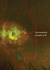
Management of central serous chorioretinopathy
Savita Madhusudhan (Liverpool, UK) outlined recommended management of central serous chorioretinopathy (CSC) (Figure 1). Central serous chorioretinopathy is the fourth most common retinopathy after AMD, diabetic retinopathy and branch retinal vein occlusion [4], with an incidence of 5.8 per 100,000 person-years [5]. A revised classification has been proposed based on simple and complex subtypes, each subtype subdivided further into three groups: primary CSC, first known episode of subretinal fluid; recurrent CSC, presence of subretinal fluid (SRF) with history or signs of resolved episodes; and resolved CSC, absence of SRF on OCT [6].
Half-dose photodynamic therapy (PDT) remains the mainstay treatment of CSC. Photodynamic therapy (verteporfin + half-dose (3mg/m²) / half-fluence (25 J/cm²) / half-time (42s)) is applicable to subfoveal / juxtafoveal CSC with / without diffuse decompensation of the retinal pigment epithelium. Approximately 80–85% of eyes with chronic CSC treated with PDT show complete resolution of SRF between 6–12 weeks, with resolution of macular pigment epithelium detachment (PED) in 78% of eyes vs 38% spontaneous resolution [7]. High-density subthreshold micropulse diose laser, targeted on abnormal hyperfluorescent areas on fluorescein angiography / indocyanine green angiography, can achieve resolution of SRF in up to 75% of eyes, with the benefit of no visible laser burns, minimal collateral damage, and safe for subfoveal leaks. Available evidence does not support the use of topical non-steroidal anti-inflammatory drugs for the treatment of CSC [8]. A recently published treatment guideline recommends that if PDT is unavailable, chronic CSC with focal, non-central leakage on angiography may be treated using conventional laser photocoagulation [9]. Also, CSC with concurrent macular neovascularisation should be treated with half-dose / half-fluence PDT and / or intravitreal anti-VEGF therapy.
Cystoid macular oedema
Richard Gale (York and Scarborough, UK) discussed how to approach unexplained cystoid macular oedema (CMO). Symptoms of CMO include blurred central vision, metamorphopsia, decreased contrast sensitivity and micropsia. Postoperative CMO is an important complication following intraocular surgery [10]. It has been shown that spectral domain OCT can be used to differentiate pseudophakic CMO (PCMO) from DMO, by an automated model as well as by trained masked readers [11]. Munk, et al. observed that PCMO usually presents with a central MO pattern and intraretinal cystoid fluid accumulation in the central millimetres and the inner Early Treatment Diabetic Retinopathy Study (ETDRS)-subfields [11]. Rabina and colleagues studied the effects of PCMO on retinal nerve fibre layer (RNFL) thickness before and after treatment using OCT, concluding that peripapillary RNFL thickness may serve as an additional parameter for diagnosis and follow-up of PCMO [12].
Clinicians should determine the underlying cause of the CMO and tailor therapy to aetiology, noted Mr Gale. Commonly reported treatments for refractory PCMO include steroid and biological-based therapies. An interventional case series report found that treatment with dexamethasone intravitreal implant (Ozurdex, AbbVie) achieved a sustained effect over up to six months in patients with chronic CMO after uncomplicated vitrectomy for epiretinal gliosis or phacoemulsification or secondary to non-infectious endogenous uveitis [13]. A meta-analysis published in 2023 confirmed favourable visual prognosis and anatomical improvement in patients with uveitic macular oedema after receiving single-dose intravitreal dexamethasone implant [14].
Sickle cell maculopathy
James Talks (Newcastle, UK) discussed sickle cell maculopathy. The prevalence of sickle cell maculopathy has been reported as 45.6% in a sickle cell disease population and increases with age [15]. Sickle cell maculopathy can affect the vision and OCT map is useful for detection, and OCT-A can detect change not seen on the OCT map. Mr Talks commented that more work is required on correlation to systemic factors.
Proliferative sickle cell retinopathy (SCR) is the most common ophthalmic manifestation of sickle cell disease and can result in significant sight loss [16]. There are five stages of SCR in the Goldberg classification: stage I, peripheral arterial occlusion; stage II, peripheral arteriovenous anastomoses; stage III, neovascular and fibrous proliferation; stage IV, vitreous haemorrhage; stage V, retinal detachment. Laser treatment is an effective option for stage 3 SCR, shown to reduce the risk of visual loss and protective against the development of vitreous haemorrhage [17]. Stage 3 and 4 SCR can be managed with intravitreal anti-VEGF therapy, and the main therapeutic intervention for stage 4 and 5 SCR is vitrectomy [16].
Ultra-widefield imaging
Tariq Aslam (Manchester, UK) reviewed the application of ultra-widefield (UWF) OCT angiography for rapid, non-invasive and reliable imaging that can often negate or avoid the need for fluorescein angiography. Mr Aslam noted broad consensus on the utility of widefield OCT angiography for detection of retinal neovascularisation in eyes with proliferative diabetic retinopathy [18].
The role of UWF imaging in myopia was explored by Ali Erginay (Paris, France) who observed that high myopic eyes present unique anatomical and pathological challenges. Ultra-widefield imaging, particularly when combined with OCT, helps in evaluating the extent and morphology of staphylomas, peripheral retinal degenerations such as lattice degeneration, retinal tears, and holes, which are often missed with conventional fundus imaging. In high myopia, with its high risk of vision-threatening complications, UWF imaging offers a comprehensive and efficient approach for early detection, precise documentation and effective monitoring. Its integration into routine clinical practice enhances the ability to manage high myopic patients proactively and more effectively, observed Erginay.
What ophthalmologists need to know about GLP-1 RA therapy
Glucagon-like peptide-1 receptor agonists (GLP-1 RAs) are an established, effective therapeutic class of glucose lowering drugs. Key findings of diabetic retinopathy (DR) complications from the SUSTAIN clinical trial programme evaluating semaglutide, a GLP-1 analogue, for the treatment of type 2 diabetes were: increased risk of DR complications consistent with early worsening of pre-existing DR, secondary to initial, rapid improvement in glycaemic control [19,20]; in a post-hoc mediation analysis, DR complications were observed in patients with pre-existing DR and attributable to the magnitude and rapidity of reduction in HbA1c in the first 16 weeks of treatment [19].
Marc Evans (Cardiff, UK) discussed the development of multidisciplinary expert consensus recommendations to facilitate optimum management of GLP-1 RAs in people with type 2 diabetes and obesity, with a focus on those at risk of ocular complications. Those that are at higher risk of a retinal event are likely to benefit the most from GLP-1 RAs due to being at higher risk of diabetes-related endpoints, and improved glucose control may reduce new onset retinopathy, explained Mr Evans. GLP-1 RAs may result in retinopathy progression in susceptible individuals, through early worsening which becomes apparent within three to six months and may resolve within 24 months, he added. It is recommended that people with diabetes prescribed a GLP-1 RA should have a retinal screen within the previous 12 months, particularly those with long duration diabetes (>10 years), poor glucose control (HbA1c >10%), or established retinopathy (pre-proliferative / proliferative).
Mr Evans commented that when prescribing GLP-1 RA in people with sight loss in one eye, prior history of non-arteritic anterior ischemic optic neuropathy (NAION) or risk factors, caution is advised [21]. He explained that he follows a staged glucose reduction approach in patients at risk of retinopathy worsening, initiating GLP-1 RA treatment with a low starting dose and maintaining treatment at a lower dose for longer.

Figure 2: Silicone oil retention sutures in a case of traumatic aniridia, aphakia and retinal detachment (four weeks post retinal detachment repair). Source: Courtesy of Roxane J. Hillier, Newcastle Eye Centre, Royal Victoria Infirmary, Newcastle upon Tyne.
The vitreoretinal surgeon and ocular trauma
Roxane Hillier (Newcastle, UK) discussed the role of the vitreoretinal surgeon in ocular trauma and demonstrated outcomes that may be achieved with contemporary devices and techniques in patients with open globe injuries (OGIs) (see Figure 2).
Timing is important when considering involvement of the vitreoretinal surgeon in cases of OGI, explained Ms Hillier. Once primary repair has been undertaken, there exists a ‘sweet spot’ between risk of acute intraoperative complications at the time of the secondary surgery (wound instability, tissue prolapse and expulsive suprachoroidal haemorrhage) versus onset of proliferative vitreoretinopathy (PVR). General ophthalmologists should inform their vitreoretinal colleagues of any repaired OGI case with posterior segment involvement early, preferably within the first few days and earlier for cases of intraocular foreign body, appositional ‘kissing’ choroidals, or infective endophthalmitis. Multiple sources suggest that the ideal interval to vitrectomy is within two weeks [22,23]. Ms Hillier said that her personal preference is vitreoretinal review at 7–10 days post primary repair, when the globe tends to be more robust and the eye has ‘declared itself’ in terms of visual acuity, intraocular pressure (IOP) and infection.
In the meantime, successful secondary surgical reconstruction is facilitated by high-quality primary closure, CT scan where bony injury or intraocular foreign body is suspected, close monitoring for infection, aggressive management of raised IOP, and regular ultrasound monitoring of the posterior segment. Multiple factors influence decision-making on secondary vitreoretinal intervention. Ms Hillier emphasised that no light perception (NLP) vison in the early days after injury is not a hard contraindication to secondary surgery, while posterior segment disorganisation is a negative prognostic factor, especially in the context of NLP vision [24].
In patients with ocular trauma, general ophthalmologists should involve vitreoretinal colleagues early and actively monitor for and treat complications in the interim. Late referral for established PVR retinal detachment is a lost opportunity, noted Ms Hillier. The decision to proceed with vitreoretinal surgery in severe OGI is complex, individualised and requires thorough patient counselling, concluded Ms Hillier, adding that excellent outcomes are possible with care, prompt attention and skill.
References
1. https://www.england.nhs.uk/greenernhs/
wp-content/uploads/sites/51/2022/07/B1728
-delivering-a-net-zero-nhs-july-2022.pdf
2. Malcolm J, Dodd A, Shaikh M, et al. Reducing the carbon footprint of cataract surgery: co-creating solutions with a departmental Delphi process. Eye (Lond) 2024;38(7):1349–54.
3. https://www.eyesustain.org/
member-societies/nog
4. Wang M, Munch IC, Hasler PW, et al. Central serous chorioretinopathy. Acta Ophthalmol 2008;86(2):126–45.
5. Kitzmann AS, Pulido JS, Diehl NN, et al. The incidence of central serous chorioretinopathy in Olmsted County, Minnesota, 1980-2002. Ophthalmology 2008;115(1):169–73.
6. Chhablani J, Cohen FB; Central Serous Chorioretinopathy International Group. Multimodal imaging-based central serous chorioretinopathy classification. Ophthalmol Retina 2020;4(11):1043–6.
7. Arif F, Pryds A, Larsen M. Isolated pigment epithelium detachment: evidence for relation to central serous chorioretinopathy and effect of photodynamic therapy. Acta Ophthalmol 2018;96(8):821–7.
8. Larsson JME, Boberg-Ans LC, Vangsted A, et al. Topical non-steroidal anti-inflammatory drugs for central serous chorioretinopathy: A systematic review and meta-analysis. Acta Ophthalmol 2024;102(3):274–84.
9. Feenstra HMA, van Dijk EHC, Cheung CMG, et al. Central serous chorioretinopathy: An evidence-based treatment guideline. Prog Retin Eye Res 2024;101:101236.
10. Bunjo LJ, Bacchi S, Pietris J, Chan WO. Current management options for the treatment of refractory postoperative cystoid macular edema: A systematic review. Surv Ophthalmol 2024;69(4):606–21.
11. Munk MR, Jampol LM, Simader C, et al. Differentiation of diabetic macular edema from pseudophakic cystoid macular edema by spectral-domain optical coherence tomography. Invest Ophthalmol Vis Sci 2015;56(11):6724–33.
12. Rabina G, Shtayer C, Shemesh R, et al. Peripapillary retinal nerve fiber layer thickness as a diagnostic tool for pseudophakic cystoid macular edema. Eur J Ophthalmol 2024;34(2):534–40.
13. Garweg JG, Baglivo E, Freiberg FJ, et al. Response of postoperative and chronic uveitic cystoid macular edema to a dexamethasone-based intravitreal implant (Ozurdex). J Ocul Pharmacol Ther 2016;32(7):442–50.
14. Fan S, Shi XY, Zhao CF, et al. Efficacy and safety of single-dose intravitreal dexamethasone implant in non-infectious uveitic macular edema: A systematic review and meta-analysis. Front Med (Lausanne) 2023;10:1126724.
15. Leitão Guerra RL, Leitão Guerra CL, Bastos MG, et al. Sickle cell retinopathy: What we now understand using optical coherence tomography angiography. A systematic review. Blood Rev 2019;35:32–42.
16. Dinah C, Balaskas K, Greystoke B, et al. Sickle Eye Project: a cross-sectional, non-interventional study of the prevalence of visual impairment due to sickle cell retinopathy and maculopathy in the UK. BMJ Open 2024;14(2):e082471.
17. Myint KT, Sahoo S, Thein AW, et al. Laser therapy for retinopathy in sickle cell disease. Cochrane Database Syst Rev 2015;2015:CD010790.
18. Drira I, Noor M, Stone A, et al. Comparison of widefield OCT angiography features between severe non-proliferative and proliferative diabetic retinopathy. Ophthalmol Ther 2024;13(3):831–49.
19. Vilsbøll T, Bain SC, Leiter LA, et al. Semaglutide, reduction in glycated haemoglobin and the risk of diabetic retinopathy. Diabetes Obes Metab 2018;20(4):889–97.
20. Bethel MA, Diaz R, Castellana N, et al. Change and diabetic retinopathy during GLP-1 receptor agonist cardiovascular outcome trials: a meta-analysis and meta-regression. Diabetes Care 2021;44(1):290–6.
21. Cai CX, Hribar M, Baxter S, et al. Semaglutide and nonarteritic anterior ischemic optic neuropathy. JAMA Ophthalmol 2025;143(4):304–14.
22. Perez EA, Scott NL, Russell JF. Improved visual outcomes after severe open-globe injuries associated with perioperative vitreoretinal evaluation. Ophthalmol Retina 2023;7(9):771–8.
23. Phillips HH, Blegen Iv HJ, Anthony C, et al. Pars plana vitrectomy following traumatic ocular injury and initial globe repair: a retrospective analysis of clinical outcomes. Mil Med 2021;186(Suppl1):491–5.
24. Sherif NA, Hoyek S, Wai K, et al. Recovery of vision in open globe injury patients with initial no light perception vision. Ophthalmol Retina 2024;8(7):617–23.
[All links last accessed June 2025]
Declaration of competing interests: The author has provided consultancy services to Bayer AG, DORC International B.V., Johnson & Johnson Vision Care, Inc., Roche Products Ltd and Thea Pharmaceuticals Ltd.