We asked the next generation of healthcare scientists – students of the BSc Healthcare Science Ophthalmic Imaging degree – about their experiences, insights and aspirations, painting a picture of how ophthalmic imaging is evolving.

Rosalyn Painter (RP): I’m here today with two students from the very first cohort of the ophthalmic imaging degree run by the University of Gloucestershire. Helen Hobman and Andy Jones are set to graduate this year as Ophthalmic Healthcare Science Practitioners (HSPs). It’s exciting to hear their thoughts on this world-first programme and what it means for their future development and the development of the ophthalmic imaging profession.
1. What inspired you to pursue a degree in ophthalmic imaging, and how do you see your role evolving within the wider field of eyecare?
Helen Hobman (HH): I was inspired by the collaboration between advancing technology and the delivery of high-quality care to our patients. Ophthalmic imaging helps to visualise the structures of the eye, which is a valuable contribution to helping manage and diagnose ophthalmic diseases. I see my role as an HSP as a key role in the multidisciplinary team, helping to interpret findings and assist in decision making regarding the next steps for imaging.
Andy Jones (AJ): I never thought I’d see the day when university was more than just a distant dream, especially with no A-levels to my name. But when this course came my way, I was beyond excited to prove to myself and become the best healthcare professional I could be, especially in the world of ophthalmic imaging. During my degree, I've had the chance to teach new starters at my hospital, Manchester Royal Eye Hospital (MREH), and I’ve discovered a true passion for teaching. I’m convinced I’ve found my calling. Once I finish this degree, I’m aiming for a role that allows me to grow in this area both for myself and, most importantly, the MREH imaging department.
2. During your training so far, what imaging techniques or technologies have you found most fascinating, and why?
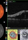
HH: In fairness I thought it would be a technique that I have not previously been exposed to, but after the three years I have to say it is optical coherence tomography (OCT) and OCT angiography. This non-invasive technique has revolutionised ophthalmic clinics and how we detect and monitor disease, making the process more patient friendly. Having the ability to document and revisit imaging to detect subtle changes allows for second opinions in MDT meetings. This technology has enabled centres for disease control [SV1.1]and virtual acquisition clinics to exist and aid in the backlog in ophthalmology.
AJ: The use of anterior-segment photography has really captured my eye (yes, pun intended!). First, it's incredible to see the stunning images it produces, and second, learning the techniques like using the slit-lamp to capture these beautiful photos has been an absolute thrill.
3. Can you describe a moment in your studies where imaging made a critical difference to a patient’s diagnosis or care?
HH: I have been able to able to apply my knowledge on numerous occasions, however one patient springs to mind, from a virtual acquisition clinic for naevus patients who are either new or follow-up patients. The patient had already waited a long time from referral, and due to the teaching around the MOLES scoring system, I saw that orange pigment was present and it was larger in size. We were unable to identify if growth was evident due to the patient being a new referral, and subretinal fluid was present. With this information it was clear the patient would need a referral to the local oncology unit.
I wanted to ensure that this patient’s referral and any additional imaging was completed, so I sought the help of the doctor on call to complete a B-Scan ultrasound as I know this is a complementary image to help assess the lesion. Being aware of the patient pathways is crucial, as this enabled me to email the reviewing consultant team to review the images earlier and the referral was completed first thing on the next working day.
AJ: Learning about retinal detachment during my time in the diagnostic clinic gave me the insight and more importantly, the confidence to recognise when a patient needs urgent surgical attention. Without that knowledge, a patient might have gone home, planning to “get it checked” in a week or two… or three, by which point the vision loss could have been permanent. It's a powerful reminder that this degree isn't just academic; it's directly improving patient care through my sharper understanding of ophthalmic diagnostics.
4. How do you think emerging technologies will change the practice of ophthalmic imaging in the next 5–10 years?
HH: AI is essential in the development of ophthalmic imaging, but caution must be taken that we do not deskill as practitioners and become reliant on automated image analyses or detection of disease. AI should be a complementary tool in patient care. Governance is a key element around AI and a very complexed area to ensure the public build trust with developing AI technologies. Additionally, I believe AI will help ophthalmic HSPs develop confidence and skillsets, as well as potentially speeding up clinical diagnosing and monitoring appointments.
AJ: MREH has already done a few studies to see if AI can detect eye conditions from just an OCT which were surprisingly accurate, even with just a sample size of 1000 patients. As AI continues to advance and more patients are added to the database, its accuracy will only improve. However, AI still has a ‘black box’ problem as we don’t always understand how it reaches its conclusions. It’s best when humans remain in the driver’s seat, using AI as a tool to work in harmony rather than letting it take over.
5. In your experience so far, what skills beyond technical knowledge are most important for success in ophthalmic imaging?
HH: I strongly believe it is a multifactorial approach to be able to deliver effective patient-centred care, which needs to be adapted for each situation to ensure individualise care is given – education, teamwork, communication, patient interaction, alongside reflection. Advocacy for staff and patients is also essential to ensure that standards are delivered in accordance with the Good Scientific Practice domains, this enables the service to deliver and emphasise care for our patients.
AJ: Ongoing training and establishing a recognised, registered board for all ophthalmic imaging departments, like what we see in ophthalmology or nursing, will help the field grow and thrive. Ophthalmic imaging is a true asset to the NHS, and it’s a gift that deserves to be recognised and supported as it continues to expand.
6. What challenges have you encountered when learning complex imaging procedures, and how have you overcome them?
HH: A major challenge for me, which is part of the portfolio, was around developing a skillset with various equipment. To ensure I was able to develop and understand what was involved in the imaging techniques, I utilised the block weeks and all the time available. Equipment comes in from all different manufactures, and using this time to speak to the experts and other students who have experience allowed me to practice skills in a controlled safe environment. I also participated in additional courses outside of university such as the Haag-Streit slit-lamp course which runs over two days – this enabled me to underpin knowledge with real-life experiences. Asking questions was key in helping me to develop my knowledge, as well as additional reading.
AJ: Through ongoing education from my university course, I’ve gained a better understanding of patients' vision limitations, allowing me to assess what they can or can't see. This helps me approach procedures with more confidence and ease. For example, high-myopic patients can be tricky, but a simple adjustment, like switching or changing the scan, can make a world of difference, making the process smoother for both the patient and myself, ultimately improving patient care.
7. If you could improve or invent one tool or technology to support ophthalmic imaging, what would it be?
HH: Handheld equipment would need to be improved for me to ensure all patients with mobility issues have the same access to imaging. I get really frustrated when we cannot obtain images for patients or be able to book them in the virtual acquisition clinics, which often run more to time than face-to-face appointments. The handheld equipment needs to be lightweight to ensure use for the practitioner is easy and does not cause any strain.
AJ: Imagine a dilation drop that only lasts for 30 minutes, allowing patients to drive themselves to appointments, or an oral or topical drop that can reverse dilation, bringing the pupil back to a safe size for driving. This would be a game-changer for patient convenience and independence.
8. How do you think the role of ophthalmic imagers could be better integrated into multidisciplinary eyecare teams in the future?
HH: Job roles need to be standardised across the country, allowing for senior managers to better understand the capabilities and skills of their workforce, and to help bridge the gap between ophthalmologists which are experiencing shortfalls in recruitment. Structured career development and registration to uphold standards and accountability. Inclusion in multidisciplinary team meetings is essential to develop learning, and to be included in interpretation of images to help reduce the backlogs.
AJ: Just as ophthalmologists assist doctors in their clinics, I believe ophthalmic imagers can play a similar role working alongside ophthalmologists to share the load. By handling the simpler cases, we can free up the doctors and ophthalmologists to focus on the more complex ones, improving efficiency and patient care across the board. RP: Thank you for sharing these fascinating insights with our readership. On a personal note, I’m looking forward to hearing where you both decide to take your careers, and congratulations on being pioneers in your field.
I hope this article has been informative and inspiring. If you or someone in your team would benefit from undertaking this degree, please get in contact with the apprenticeship team at the university of Gloucestershire: futureapprentices@glos.ac.uk