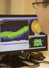
From monitoring patients with heart disease to improving the early diagnosis of cancer, artificial intelligence (AI) using deep learning techniques is already employed in many different healthcare specialties [1]. In the eyecare field, AI technologies have been used to analyse retinal images for automatic detection of diabetic eye disease, age-related macular degeneration, glaucoma, and systemic vascular and metabolic diseases [2].
Such technologies have the potential to improve the diagnosis and management of ocular and systemic diseases manifesting in the eye by providing valuable, fast clinical decision-making support to eyecare professionals. But are there any factors we need to consider to ensure these technologies are truly going to bring these benefits to our patients?
Debates surrounding the ethics of AI are widespread in the media currently. Much of this focuses on Generative AI, a subset of AI which is used to create plausible content including images, text, audio and video. When an AI-generated picture can win an art prize or an AI chatbot can pass a law school exam, it makes sense that we would want to examine how we got here and where we might be going [3,4].

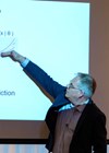
Figure 1: The AI-generated artwork “Théâtre D’opéra Spatial”, generated by Jason M Allen
on Midjourney, won first prize in the digital art category at the Colorado State Fair.
Much of the debate surrounding these high-profile AI tools scrutinises the way in which the algorithms were trained. Deep learning algorithms require large volumes of data in order to learn how to perform a specific task. In the case of an image-generating AI such as Midjourney, the AI used to create the prize-winning digital art in Figure 1, this training dataset took the form of hundreds of millions of images mined from the internet. Included in these images were original creations from artists past and present, raising questions about the fairness of using copyrighted creative work to train an AI algorithm and what it means for artists’ jobs and the nature of art itself.
When it comes to the development of AI for retinal image analysis a different discussion emerges. Firstly, unlike AI art-generating tools, retinal image analysis technologies are designed to support but not to replace the role of the eyecare practitioner. Furthermore, in contrast to the public domain art images used to train Midjourney, the retinal image datasets used to train deep learning algorithms are subject to stringent governance and ethical standards.
That does not, however, mean that these retinal image datasets are without issue, and a deep learning system is only ever as good as the data is has been trained on.
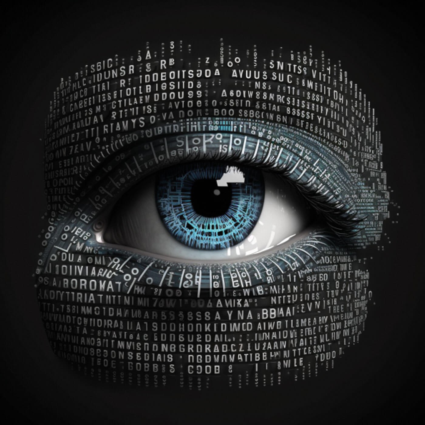
Figure 2: An AI-generated image of an eye created in Midjourney.
Some retinal image datasets contain relatively homogenous populations with limited ethnic diversity within them. This could lead to shortcomings in how well a deep learning system performs on those from ethnic minority groups. Racial bias has been observed in many deep learning systems in the wider AI field, such as the prediction of crime hot spots, facial recognition, or job application screening [5-7]. Care should be taken to ensure similar biases do not arise in retinal diagnostic tools, as this could have serious implications for health equity.
Most widely used retinal image datasets have been acquired either in a research or hospital-based setting. Research-acquired retinal image datasets often only contain a single image capture session and so represent a snapshot of only one point in time. Hospital-acquired datasets from ophthalmology services often contain multiple image capture sessions which provide more information about how the retina changes over time in the presence of disease. However, the retinal images included in these datasets generally come from patients who already have a suspected or confirmed diagnosis of eye disease and therefore do not capture the early stages of disease which are essential to develop early monitoring and prediction technologies.
So how do we create a retinal image dataset that captures the early stages of disease? Or better yet, how do we capture the patient in the pre-diagnostic phase, allowing for the discovery of new biomarkers and earlier diagnosis?
Community optometry in Scotland provides NHS-funded eye examinations for all, which has included digital retinal photography for patients aged 60 plus since 2008. Eye examination uptake has increased in Scotland since the introduction of this funding model which removed the cost barrier to accessing primary eye care, and consequently optometrists in Scotland now capture millions of retinal images every year. Many optometry practices have been capturing retinal images for a decade or more. Currently, those images sit within each individual practice, contributing to the individual care of each patient within the practice, but remaining untapped as a potential resource for eye care research. Harnessing the power of these community-acquired retinal images is the goal of the Scottish Collaborative Optometry-Ophthalmology Network e-research (SCONe).
SCONe aims to transfer retinal images captured by community optometrists in Scotland to the National Safe Haven, a secure NHS storage facility run by Public Health Scotland. Once in the Safe Haven, the images are linked to routinely-collected NHS data and pseudonymised so that no personal identifiers are visible, creating a rich resource for innovation in research, healthcare and education. As community optometrists in Scotland provide both routine eye examinations and acute eyecare services, the retinal images captured in this setting contain patients at all stages of the disease process including those who are healthy, pre-diagnostic, early, moderate and in the late stages of disease. By investigating the pre-diagnostic stage, the discovery of novel biomarkers enabling earlier detection of disease can be realised. The rich dataset created by the linked NHS data will create opportunity to discover retinal manifestations of systemic diseases. And the longitudinal nature of the dataset will enable researchers to travel through time and discover how the retina changes in ocular and systemic diseases.
The creation of the SCONe research resource is subject to stringent governance protocols. Scotland has a rich history in the use of non-consented healthcare data for public benefit as governed by the Charter for National Safe Havens (2015) [8]. At every stage of the project, SCONe has taken steps to ensure data privacy and security are upheld at all times, details of which can be found in the project’s data privacy statement [9]. SCONe received approval from NHS Scotland’s Public Benefit and Privacy Panel (PBPP), the highest governing body in NHS Scotland, in October 2021.
Figure 3: An AI-generated image of an eye created in Midjourney.
Following the completion of the relevant governance and ethical approvals, SCONe commenced transferring retinal images in its Proof of Concept phase in early 2022. The SCONe team, which consists of optometrists, ophthalmologists, data scientists and IT experts, has since gathered tens of thousands of retinal images from Scottish community optometry practices. Over 30,000 retinal images have been delivered to the National Safe Haven and successfully linked to routinely-collected NHS data, with a further ~100,000 images extracted from practices which are currently undergoing processing for linkage. This means that SCONe will soon be one of the world’s largest repositories of longitudinal community-acquired retinal images.
As SCONe moves beyond its Proof of Concept phase, the team has begun working towards Scotland-wide coverage. The SCONe project aims to create a dataset which is truly representative of primary care patients, with an ethnicity, age and socio-economic range reflective of the diversity in Scotland’s population. Such a dataset will enable robust validation of new AI technologies that are developed to help eyecare practitioners to diagnose early, monitor and manage patients with confidence, and improve patient outcomes.
With the Scottish Government expressing their support for this “globally important study”, optometry practices across Scotland are encouraged to take their place as innovators of eyecare research by contributing retinal images to the SCONe research resource [10]. Optometry practices in Scotland can register their interest in joining via the SCONe website, or by emailing scone@ed.ac.uk
References
1. Yu KH, Beam AL. I. Kohane IS, Artificial intelligence in healthcare. Nat Biomed Eng 2018;2(10):719-31.
2. McNeil R. Coming to terms with AI. Eye News 2019;26(2):6-14.
3. Roose K. An A.I.-Generated Picture Won an Art Prize. Artists Aren’t Happy. The New York Times
https://www.nytimes.com/2022/09/02/
technology/ai-artificial-intelligence-artists.html
4. Sloan K. ChatGPT passes law school exams despite ‘mediocre’ performance. Reuters
https://www.reuters.com/legal/
transactional/chatgpt-passes-law-school
-exams-despite-mediocre
-performance-2023-01-25/
5. Amnesty International UK. Call to Ban Facial Recognition Technology that amplifies racist policing.
https://www.amnesty.org.uk/press-releases/
call-ban-facial-recognition-technology
-amplifies-racist-policing
6. Marsh S. “UK police use of computer programs to predict crime sparks discrimination warning”. The Guardian
https://www.theguardian.com/uk-news/
2019/feb/03/police-risk-racial-profiling-by
-using-data-to-predict-reoffenders-report-warns
7. Milmo D. UK data watchdog investigates whether AI systems show racial bias. The Guardian
https://www.theguardian.com/technology/
2022/jul/14/uk-data-watchdog-investigates
-whether-ai-systems-show-racial-bias
8. Department of Health and Social Care. Charter for Safe Havens in Scotland: Handling Unconsented Data from National Health Service Patient Records to Support Research and Statistics.
https://www.gov.scot/publications/charter-safe
-havens-scotland-handling-unconsented-data
-national-health-service-patient-records
-support-research-statistics/
9. SCONe. Data Privacy Statement. The University of Edinburgh.
https://www.ed.ac.uk/clinical-sciences/
ophthalmology/scone/data-privacy-statement
10. Directorate of Primary Care. PCA(O)2022(07).
https://www.sehd.scot.nhs.uk/
pca/PCA2022(O)07.pdf
11. European Parliament. What is artificial intelligence and how is it used?
https://www.europarl.europa.eu/news/
en/headlines/society/20200827STO85804/
what-is-artificial-intelligence-and-how-is-it-used
12. Pead E, Megaw R, Cameron J, et al. Automated detection of age-related macular degeneration in colour fundus photography: a systematic review. Surv Ophthalmol 2019;64(4):498-11.
13. Scotland GS, McNamee P, Fleming AD, et al. Costs and consequences of automated algorithms versus manual grading for the detection of referable diabetic retinopathy. Br J Ophthalmol 2010;94(6):712-9.
14. National Institute for Health and Care Excellence (NICE). AI technologies for detecting diabetic retinopathy.
https://www.nice.org.uk/advice/mib265/
resources/ai-technologies-for-detecting
-diabetic-retinopathy-pdf-2285965755558853
15. Diabetes UK. Diabetes diagnoses double in the last 15 years.
https://www.diabetes.org.uk/about_us/news/
diabetes-diagnoses-doubled-prevalence-2021
[All links last accessed December 2022].
Declaration of competing interests: None declared.
COMMENTS ARE WELCOME