The authors remind us of the revolutionary impact gases have had on retinal surgery.
In 1938, Rosengren attempted to improve his retina repair surgical outcomes [1]. He discovered that suturing the retina or pressing externally on the sclera were not the only methods that “secure the coalescence of the retina and bulb capsule”. Instead, he introduced the concept of internal gas tamponade, which is now the primary method used following vitrectomy to ensure retinal re-attachment.
In 1970, Machemer et al. introduced the pars plana approach to vitrectomy that has since revolutionised our current practice [2]. Today, three vitreous substitutes are commonly used: intraocular gases, silicone oil and perfluorocarbon liquid. In this article, we aim to explain the different properties, the common indications, the contraindications and the complications of the various intraocular gas vitreous substitutes.
Four intraocular gases are most frequently used: air, sulfur hexafluoride (SF6), perfluoroethane (C2F6), and perfluoropropane (C3F8). The gas’ high surface tension allows the retina to re-attach by giving the retinal pigment epithelium time to pump out the residual subretinal fluid. An important physical property that affects postoperative management is buoyancy. Buoyancy is the upward force exerted by the gas against the retina. The apex of the bubble can be directed against the retinal tear to create the ideal tamponade by positioning the head of the patient. This will then allow the retinopexy to take effect whilst maintaining the tamponade of the retina to the retinal pigment epithelium.
Every intraocular gas inserted into the vitreous has three phases: expansion, equilibrium, and dissolution [3]. Air doesn’t expand, with its duration being five to ten days. SF6 duration is 14 days, peak expansion is one to two days, and non-expansile concentration is 20%. C2F6 duration is four weeks, peak expansion is 36-60 hours, and non-expansile concentration is 16%. C3F8 duration is between six to eight weeks, peak expansion is 72-96 hours, and non-expansile concentration is 12% [3]. The most rapid expansion rate is achieved in the first six to eight hours after the placement of gas.
Use of intraocular gases
Intraocular gases can be used in two settings. In cases of pars plana vitrectomy for retinal detachment, either an expansile concentration or non-expansile concentration is used depending on the amount of residual subretinal fluid. In cases of pneumatic retinopexy where the vitreous is preserved, an expansile concentration is used to create a focal tamponade point over the break.
In cases of pneumatic retinopexy, a thorough examination using indirect ophthalmoscopy should be done to ensure continued perfusion of the central retinal artery, and an anterior chamber paracentesis should be employed if the intraocular pressure is thought to be high.
In cases of pars plana vitrectomy, the subretinal fluid is evacuated either through the retinal tear or a surgical retinotomy. Subsequently, a fluid air exchange is done by passive or active aspiration. Passive aspiration can be done using a flute needle and active aspiration can be done utilising an extrusion needle or the vitrectomy cutter. An appropriate gas is then inserted through the port. The choice of which gas and what concentration to be used is multifactorial, depending on the ability of the patient to posture afterwards, the position of the tear and the amount of subretinal remaining. Traditional thinking is to use SF6 for superior breaks, C2F6 for equatorial breaks and C3F8 for inferior breaks (Figure 1). This doctrine has somewhat changed in modern days with the combined technique of using a scleral buckle and vitrectomy for inferior breaks with an expansile shorter-acting gas.
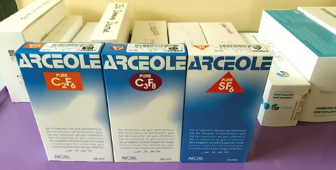
Figure 1: Commonly used gases in vitreoretinal surgery.
More recently, the use of 23 or 25- gauge vitrectomy has meant that the procedure is primarily sutureless and the integrity of the sclerotomy wound is preserved without the need of closing it. Injecting the gas is done through a sterile filter to reduce the risk of infection. The 50ml syringe is flushed several times to prevent decontamination of the pure gas with air, altering the concentration. The ideal concentration of gas is then chosen. Gas is then injected through the infusion site whilst the trocars are in situ. They are then removed and the integrity of the wounds are checked and the pressure of the eye manually assessed.
Below, we run through some of the indications of intraocular gases, the contraindications and the complications.
Indications
Macular hole surgery involves a posterior vitrectomy, followed by induction of a posterior vitreous detachment, an internal limiting membrane peel and finally insertion of intraocular gases followed by posturing postoperatively. Kim et al. found that the results of using SF6 or C3F8 were similar in terms of visual outcomes [4]. It is still debatable how long the patient should posture following a macular hole surgery. The size of the macular hole alters gas selection with smaller holes under 400 microns requiring a shorter-acting gas and larger holes requiring a longer-acting gas.
Pneumatic retinopexy is another indication to use intraocular gases. It is usually performed in superior breaks with retinal detachment. The expansile concentration is used in this case and the intraocular pressure is monitored. If the intraocular pressure is raised, then anterior chamber paracentesis can be done or, in rarer instances, vitreous aspiration, however this carries with it the risk of further retinal detachment [5]. Subretinal haemorrhage can be a devastating complication of age-related macular degeneration and lead to profound visual loss. Intraocular gases can be used to displace the blood and commonly C3F8 has been used in these instances. Intravitreal or subretinal tissue plasminogen activator (tPA) can also be injected in the same setting [6]. It should also be noted that previously intraocular gases were used to unroll giant retinal tears. This has now been replaced by using perfluorocarbon liquid in modern techniques [3].
Contraindications and complications
Three main relative contraindications to intraocular gas exist. First, air travelling should be avoided for patients with intraocular gases as it can lead to the expansion of the gases when at a high altitude. This can, in turn, can lead to a raised intraocular pressure with consequently a retinal artery occlusion and rarely ocular tissue expulsion through the previous sclerotomies. The minimal acceptable residual dose of gases is around 0.6ml to 1.0ml [7]. Similiarly diving can lead to a shrinkage of the intraocular gases, decreasing the intraocular pressure, which can lead to suprachoroidal haemorrhage and choroidal effusions [8]. Finally, nitrous oxide anaesthetic should be avoided in cases of retinal detachment as it can lead to pressure changes during the gas expansion. This can lead to an initial hypotony then followed by a hypertensive intraocular phase [9]. Therefore, any patient receiving intraocular gas should wear a wristband stating the name and concentration of gas used. This helps raise awareness of intraocular gas should a general anaesthetic be required.
Significant complications of intraocular gases are raised intraocular pressure, gas cataract, and migration of intraocular gases through the subconjunctival space, subretinal space or anterior chamber in aphakic patients. Increased intraocular pressure is usually transient and can sometimes be due to secondary angle-closure glaucoma. Gas-induced cataract or feathering of the lens surface can be reversed and disappears after the intraocular gases naturally clears. Nonetheless, intraocular gases can lead to the progression of nuclear sclerosis if already present before surgery. Finally, migration of intraocular gases to subretinal space is rare but can be seen in cases of pneumatic retinopexy. This can be uncomfortable for the patient and lubrication is advised.
TAKE HOME MESSAGE
-
The introduction of intraocular gases has revolutionised vitreoretinal surgery.
-
Different types of gases are used based on their properties.
-
Keep in mind the indications, contraindications, and complications of using them.
References
1. Rosengren B. Results of treatment of detachment of the retina with diathermy and injection of air into the vitreous. Acta Ophthalmol 1938;16:573-9.
2. Machemer R, Buettner H, Norton EW, Parel JM. Vitrectomy: a pars plana approach. Trans Am Acad Ophthalmol Otolaryngol 1971;75:813-20.
3. Mohamed S, Lai TYY. Intraocular gas in vitreoretinal surgery. HKJO 2010;14(1):8-13.
4. Kim SS, Smiddy WE, Feuer WJ, Shi W. Outcomes of sulfur hexafluoride (SF6) versus perfluoropropane (C3F8) gas tamponade for macular hole surgery. Retina 2008;28(10):1408-15.
5. Chan CK, Lin SG, Nuthi AS, Salib DM. Pneumatic retinopexy for the repair of retinal detachments: a comprehensive review (1986-2007). Surv Ophthalmol 2008;53:443-78.
6. Stroman WR, Gross JG, Taylor RJ, Rodgers FL. Current treatment strategies for submacular haemorrhage. Expert Review of Ophthalmology 2017;12(2):159-72.
7. Mills MD, Devenyi RG, Lam WC, et al. An assessment of intraocular pressure rise in patients with gas-filled eyes during simulated air flight. Ophthalmology 2001;108:40-4.
8. Butler FK Jr. Diving and hyperbaric ophthalmology. Surv Ophthalmol 1995;39:347-66.
9. Fu AD, McDonald HR, Eliott D, et al. Complications of general anesthesia using nitrous oxide in eyes with preexisting gas bubbles. Retina 2002;22:569-74.
COMMENTS ARE WELCOME