Despite the clinical weight of the field, ophthalmology was a specialty we encountered infrequently during our early medical training. In spite of limited formal exposure during undergraduate and postgraduate training, eye-related presentations were a regular feature of our day-to-day work across emergency departments, general medical wards and outpatient settings.
Resident doctors were often the first clinicians asked to review patients presenting with red eyes, visual disturbance, foreign body sensation or periorbital swelling – frequently out of hours, when senior or specialist support was not immediately available. These encounters initially felt intimidating and exposed a clear mismatch between clinical responsibility and training [1,2].
Early on, many of these consultations were accompanied by a sense of uncertainty: uncertainty about what constituted a red flag, how much examination was sufficient, and when escalation was truly necessary. Over time, however, repeated exposure and informal teaching helped us recognise that ophthalmology need not be approached as an opaque or inaccessible specialty. This article reflects the practical lessons we learned through lived clinical experience, highlighting the small number of high-yield skills that gradually transformed anxiety into confidence.
Take a structured, high-yield eye history
In our early foundation years, we often felt pressured to move quickly to examination when assessing patients with eye complaints, particularly in busy acute care settings. With experience, we learned that a structured ophthalmic history frequently provided more diagnostic clarity than examination alone, especially when specialist equipment was unavailable [3].
Clarifying the onset and progression of symptoms became central to our assessments. Sudden visual loss or severe pain immediately prompted escalation, whereas more gradual changes often suggested less acute pathology. Asking explicitly about pain proved essential, as painful eye conditions such as keratitis, uveitis, scleritis and acute angle-closure glaucoma require urgent attention [4,5]. Patients can volunteer symptoms such as flashes, floaters or a curtain-like visual defect, with these features frequently indicating retinal disease needing attention.
Contact lens use became one of the most influential questions in our history-taking. On several occasions, relatively mild symptoms in contact lens wearers masked early infectious keratitis, reinforcing the importance of a low threshold for referral [6]. Exploring a history of trauma, including minor or occupational injuries, also proved invaluable. Over time, we found that a thorough history alone often identified patients who required same-day ophthalmology review.
Visual acuity: the ophthalmic vital sign
During our early on-call shifts, visual acuity was not always prioritised. In retrospect, this often reflected an uncertainty about how to assess vision outside of clinic environments rather than a lack of understanding of its importance. However, feedback from senior colleagues and ophthalmology teams repeatedly highlighted visual acuity as the cornerstone of safe eye assessment.
With experience, we learned that consistently measuring and documenting visual acuity was one of the most transformative changes we could make to our practice. Measuring each eye separately, using the patient’s usual correction where possible, and clearly documenting testing distance improved the clarity of our referrals and facilitated more efficient triage [7,8].
The pinhole test became an invaluable learning tool. Observing improvement with pinhole often suggested refractive error and provided immediate reassurance, while lack of improvement prompted earlier escalation and avoided false reassurance. Incorporating visual acuity into every assessment gradually reduced our anxiety and provided a reliable framework for approaching unfamiliar ophthalmic presentations.
External eye examination without a slit lamp
Early in our training, the absence of specialist equipment such as slit lamps left us feeling that meaningful eye examination was not possible outside ophthalmology clinics. Over time, however, we learned that a structured pen-torch examination could still yield significant clinical information.
Careful inspection of the eyelids often revealed ptosis, swelling, erythema or focal lesions that guided diagnosis. Assessing the pattern of conjunctival injection helped us distinguish superficial irritation from deeper inflammation, while close observation of the cornea for loss of clarity, opacities or focal defects became routine. Pupil size, symmetry and reactivity were always assessed, and regular practice of the swinging torch test improved our ability to detect relative afferent pupillary defects.
Developing and repeatedly applying a consistent examination sequence reduced the likelihood of missing important signs and increased confidence when assessing patients in unfamiliar or time-pressured settings.
Recognising ophthalmic emergencies
One of the most important lessons we learned was that junior doctors are not expected to make definitive ophthalmic diagnoses, but they must be able to recognise red-flag features that require urgent specialist input. Conditions such as acute angle-closure glaucoma, infective keratitis, uveitis, orbital cellulitis, retinal detachment and central retinal artery occlusion are all sight-threatening and require same-day review [5].
Chemical eye injuries were particularly memorable learning experiences. Through both formal teaching and real clinical encounters, we learned that immediate and copious irrigation must take priority over all other assessments, including visual acuity. This exception to the usual assessment sequence was repeatedly emphasised in national guidance and reinforced the importance of decisive early action [9].
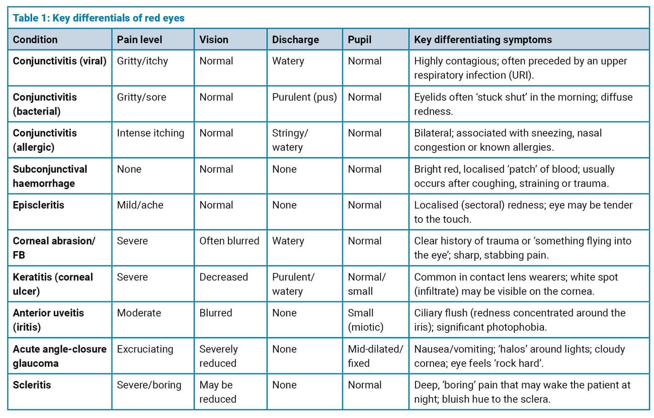
In practice, distinguishing benign from sight-threatening causes of red eye (Table 1) was something we initially found challenging, particularly during busy on-call shifts. Over time, comparing key clinical features became a useful mental framework when deciding which patients required urgent review. This comparison allows rapid differentiation between benign and sight-threatening causes [5].
Additional sight-threatening emergencies
Giant cell arteritis
Giant cell arteritis (GCA) is an important cause of acute visual loss in older adults. Suspect GCA in patients over 50 presenting with new-onset headache, scalp tenderness, jaw claudication or constitutional symptoms. Visual disturbance, diplopia or amaurosis fugax indicate ophthalmic involvement. Any suspected GCA with visual symptoms requires same-day ophthalmology referral for prompt high-dose corticosteroids. Patients without visual involvement but with systemic features should be referred urgently to rheumatology for diagnostic workup and steroid tapering.
Trauma
Traumatic ocular presentations range from superficial injuries to sight-threatening emergencies. Penetrating eye injuries and suspected globe rupture require immediate ophthalmology involvement. Signs include markedly reduced vision, irregular or peaked pupil, low intraocular pressure or visible uveal tissue. Avoid pressure on the globe and apply a rigid eye shield, review tetanus status, administer broad-spectrum antibiotics and arrange urgent CT orbit imaging without delaying specialist referral.
Chemical
Chemical eye injuries are time-critical emergencies. Irrigation takes absolute priority over all assessments, including visual acuity. Begin immediate irrigation with at least one litre of saline or until ocular surface pH normalises. Ophthalmology will not accept the case until irrigation has been completed adequately. Early aggressive management reduces the risk of limbal stem cell loss and permanent scarring.
Diplopia
Diplopia may indicate neurological or ophthalmic emergencies. An isolated third nerve palsy with pupil involvement suggests a compressive aneurysm and requires urgent neuroimaging and same-day ophthalmology and neurology input. Diplopia may also present in thyroid eye disease, myasthenia gravis and GCA.
Transient monocular visual loss
Transient monocular visual loss (amaurosis fugax) is a retinal transient ischemic attack equivalent and warrants urgent stroke team referral. Carotid stenosis, atrial fibrillation or embolic sources are common causes. Ophthalmology input is indicated when retinal pathology is suspected or diagnosis is uncertain.
Fluorescein is your friend
Fluorescein staining was initially underused in our practice, largely due to unfamiliarity and lack of confidence. With time, it became one of the most valuable investigations in acute eye assessment.
Using fluorescein allowed us to identify corneal abrasions, ulcers, herpetic dendritic lesions, dry eye patterns and signs of globe perforation more confidently [10]. We learned to always ask about contact lens use before staining, as painful eyes in contact lens wearers consistently warranted urgent ophthalmology review regardless of fluorescein findings. Taking time to explain temporary discolouration of tears and vision also improved patient reassurance and trust.

In addition to fluorescein staining, other focused assessments such as digital pressure estimation and ocular motility testing are useful adjuncts in the acute setting (Table 2).
Managing common eye presentations
Repeated exposure to common ophthalmic conditions gradually improved our confidence in managing them independently. We frequently encountered viral conjunctivitis, for example, which often required reassurance rather than intervention. Understanding when bacterial infection is more likely allows for the prescription of topical antibiotics appropriately and safely.
Similarly, chalazia and styes are best managed conservatively with warm compresses, and subconjunctival haemorrhages are typically benign and self-limiting. Recognising these patterns reduces unnecessary referrals and allows more efficient use of specialist services, while also improving patient satisfaction.
Learning and skill development
Given the limited formal exposure to ophthalmology during training, we found that actively seeking learning opportunities was essential. Shadowing eye casualty clinics, requesting bedside teaching from ophthalmology colleagues and observing orthoptists assess ocular motility were particularly valuable.
We found that digital learning resources, including e-learning modules, virtual simulations and online image libraries, helped consolidate learning and improve pattern recognition. These self-directed strategies played a key role for us in building confidence over time.
These experiences reinforced for us that confidence in ophthalmology develops incrementally. Each patient assessment, referral and discussion with senior colleagues contributed to our deeper understanding of risk and urgency. Over time, this translates into improved patient safety, clearer communication and greater professional satisfaction.
Repeated exposure to ophthalmic presentations helped us recognise patterns and normal variants, reducing stress for both us and our patients. Importantly, this confidence does not come from memorising diagnoses, but from understanding which features truly matter – pain, changes in visual acuity and red-flag symptoms – and responding appropriately. This shift allows consultations to feel more controlled and purposeful, even during busy on-call shifts.
As our experience grew, one of the most noticeable changes was not simply an increase in knowledge, but a gradual improvement in clinical judgement and confidence. Early in our training, uncertainty often led us to over-investigate or escalate prematurely, driven by a fear of missing serious pathology. While caution is appropriate, we learned to balance vigilance with reassurance, using structured assessment to guide decision-making rather than anxiety alone.
Conclusion
Ophthalmic presentations are common across all clinical environments, yet many junior doctors feel underprepared to assess them confidently. Our experience has shown that focusing on a small number of high-yield skills – structured history-taking, accurate visual acuity measurement, recognition of red flags and basic examination – can significantly improve patient safety and clinician confidence.
By sharing these experience-based lessons, we hope to reassure junior doctors that ophthalmology need not be intimidating. With gradual exposure, reflective learning and repeated practice, uncertainty can be transformed into confidence, benefiting both clinicians and patients.
References
1. Hill S, Dennick R, Amoaku W. Present and future of the undergraduate ophthalmology curriculum: a survey of UK medical schools. Int J Med Educ 2017;8:389–95.
2. Scantling-Birch Y, Naveed H, Tollemache N, et al. Is undergraduate ophthalmology teaching in the United Kingdom still fit for purpose? Eye (Lond) 2022;36(2):343–5.
3. American Academy of Ophthalmology. Basic Ophthalmology: Essentials for Medical Students. 10th ed. California, USA; American Academy of Ophthalmology; 2016.
4. AAO PPP Glaucoma Committee, Hoskins Center for Quality Eye Care. Primary angle closure disease preferred practice pattern. American Academy of Ophthalmologists 2026 [Online]:
www.aao.org/education/preferred-practice
-pattern/primary-angle-closure-disease-ppp
5. https://cks.nice.org.uk/topics/red-eye
6. AAO PPP Cornea/External Disease Committee, Hoskins Center for Quality Eye Care. Bacterial Keratitis PPP 2023. American Academy of Ophthalmology 2024 [Online]:
www.aao.org/education/preferred-practice
-pattern/bacterial-keratitis-ppp-2023
7. www.rcophth.ac.uk/wp-content/uploads/2022/
03/Curriculum-UG-RCOphth-220309.pdf
8. https://rcem.ac.uk/clinical-guidelines
9. Corbett MC, Bizrah M. Chemical Injuries of the Ocular Surface. FOCUS 2018 [online]:
www.rcophth.ac.uk/wp-content/uploads/2021/
01/College-News-FOCUS_April2018.pdf
10. Bunya VY, Mendelsohn A, Halfpenny C, et al. Removal of Corneal Foreign Bodies. EyeWiki 2026 [online]:
https://eyewiki.aao.org/Removal_of_Corneal_Foreign_Bodies
[All links last accessed July 2026]
Declaration of competing interests: None declared.