History
-
A 72-year-old male presented to his GP with a large ulcerated lesion on his right lower eyelid present for six months and enlarging in size.
-
He had an urgent referral to a tertiary centre for specialised examination.
-
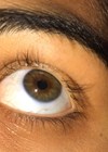
The lesion was associated with blepharitis and loss of lashes.
-
Due to the size of the tumour an exenteration was performed (Figure 1A and 1B) following a diagnostic incisional biopsy.
-
The specimen was submitted for ophthalmic histopathological assessment.
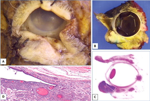
Figure 1.
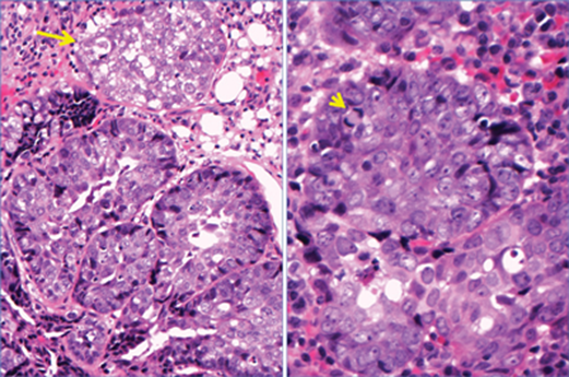
Figure 2.
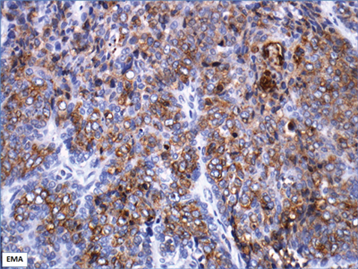
Figure 3.
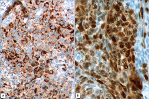
Figure 4.
Questions
-
Considering clinical information and Figure 1A and B, what clinical differential diagnoses could be raised?
-
Describe Figures 1B, C and D.
-
How can Figure 2 be described?
-
Figure 3 shows immunohistochemistry with tumour expression of epithelial marker (EMA). Figure 4 shows further two markers. Based on their positivity patterns, which markers are these anticipated to be?
-
Based on clinical features, histological and immunohistochemical findings, what is the most likely diagnosis?
Answers
1. The information of an ulcerated growing eyelid lesion associated with blepharitis and madarosis, together with the presence of a full thickness eyelid tumour raises the possibility of an infiltrative neoplasia such as squamous cell carcinoma, basal cell carcinoma, sebaceous carcinoma, etc. The presence of madarosis is helpful in narrowing down clinical suspicions.
2. Figure 1B and C shows a sagittal section of the exenteration specimen and corresponding scanned histological section demonstrating a lower eyelid full thickness tumour. Figure 1D corresponds to the white rectangle in 1C, magnifying the lower eyelid and fornix containing an infiltrative tumour.
3. The H&E histological sections show a lobulated tumour with vacuolated cytoplasm (arrow) and nuclear atypia. Note mitotic figure (arrowhead).
4. Figure 4A shows cytoplasmic expression, in areas with multiple small dots / granular appearance characteristics of adipophilin. Figure 4B shows nuclear expression of androgen receptor. 5. Sebaceous carcinoma.
COMMENTS ARE WELCOME