This site uses cookies. By continuing to browse the site you are agreeing to our use of cookies.  Read our Cookies Policy.
Close
Eye News
  • Features
    • Close
    • Features
    • Allied Professions
    • Humanitarian
    • Interviews
    • AI & Oculomics
    • Ophthalmology
    • Optometry
    • Podcast videos
    • Supplements
  • Education
    • Close
    • Education
    • Learning Curve
    • Quiz
    • Top Tips
    • Trainees
    • Medico-Legal
    • The Truth Behind The Headlines
    • Case Reports
    • Pete's Bogus Journey
  • Reviews
    • Close
    • Reviews
    • Book Reviews
    • Journal Reviews
    • What's trending?
    • Tech Reviews
    • My Top Five
    • The Culture Section
  • Events
  • News
  • Product Guide
  • Industry News
  • Contact us
    • Close
    • Contact us
    • Write for Eye News
  • Home
  • Education
  • Quiz
  • Quiz Dec/Jan 2016

Dec/Jan 2016 Quiz

1 December 2015 | EYE - Cataract, EYE - Cornea, EYE - Glaucoma, EYE - Imaging, EYE - Neuro-ophthalmology, EYE - Oculoplastic, EYE - Oncology, EYE - Orbit, EYE - Paediatrics, EYE - Pathology, EYE - Refractive, EYE - Strabismus, EYE - Vitreo-Retinal, EYE - General
Share This


History

A 40-year-old swimming instructor undergoes an enucleation for a blind painful eye. There is a history of soft contact lens wear. She has had a corneal abscess and corneal graft procedures in the past.


Questions


1. What does this immunohistochemical stain of cornea demonstrate (Figure 1)?
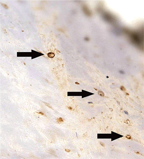
 

Figure 1.


2. What does this field including sclera and choroid show (Figure 2)?
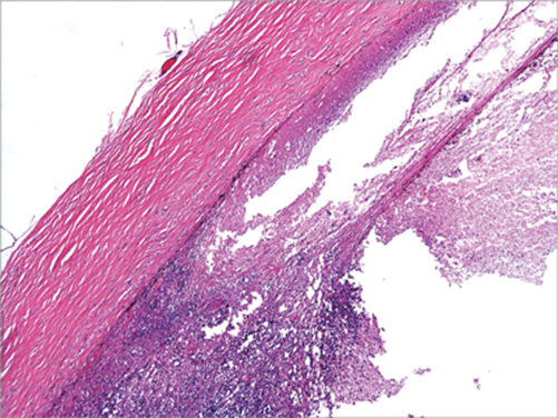
 

Figure 2.


3. What does this field including sclera and choroid show (Figure 3)?
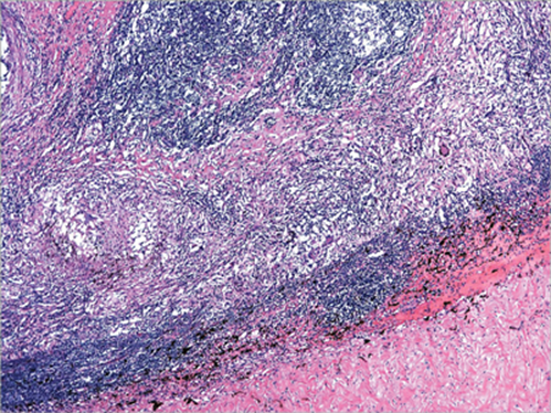
 

Figure 3.


4. What does this higher power field show (Figure 4)?
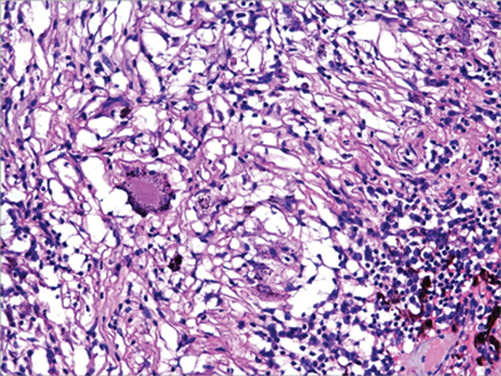
 

Figure 4.


5. What is the major clinical concern?


Answers


1. Acanthamoeba cysts.
2. Necrosis and inflammation consistent with acute endophthalmitis.
3. Granulomatous inflammation on a background of heavy lymphocytic infiltrate.
4. Multinucleate giant cells and pigment phagocytosis.
5. The granulomatous inflammation in the region of Bruch’s membrane raises the suspicion that this could be an exciting (inciting) eye in sympathetic ophthalmitis.


Discussion

This globe showed Acanthamoeba cysts within the cornea (but not the sclera or choroid), necrotising endophthalmitis with barely recognisable intraocular structures, and granulomatous inflammation which was located roughly in the region of the choroid. No other organisms were identified. The granulomata could conceivably have been part of the acute endophthalmitis process, but their location was highly suspicious of sympathetic ophthalmitis. Acanthamoeba infection may be associated with granulomatous inflammation, but endophthalmitis due to Acanthamoeba has only been reported a couple of times.


References

1. Castiblanco CP, Adelman RA. Sympathetic ophthalmia. Graefes Arch Clin Ex. Ophthalmol 2009;247:289-302.
2. Davis MJ, Packo KH, Epstein RJ, Grostern RJ, Cohen JA. Acanthamoeba endophthalmitis following penetrating keratoplasty for Acanthamoeba keratitis. Arch Ophthalmol 2010;128:505-6.


COMMENTS ARE WELCOME

Share This
Originally Published
EYE NEWS VOLUME 22 ISSUE 4 DECEMBER/JANUARY 2016
Download the full article
Download Article

Top Of Page

9 Gayfield Square, 
Edinburgh EH1 3NT, UK.

Call: +44 (0)131 557 4184
www.pinpoint-scotland.com

WEBSITE DETAILS
  • Cookie Policy
  • Data Protection Notice
  • Privacy Policy
  • Terms and Conditions
ABOUT US
  • Who we are
  • Register
  • Contact us
  • Contributors
  • Company Awards
DIGITAL ISSUES/GUIDELINES
  • Digital issues - Library
  • Supplements - Library
  • Guidelines
Accreditations
IPSO_FLAG_TEAL 2025.png cpdcertified.png

Pinpoint Scotland Ltd (Registered in Scotland No. SC068684) | © 2025 - Website by Gecko Agency