History
-
A 30-year-old male presents with a phthisical left eye and undergoes enucleation. He has some lesions in his right eye that are under ophthalmic surveillance.
-
Figure 1 is a low power of the enucleation.
-
Figures 2, 3 and 4 are higher power shots of the arrowed lesion in Figure 1.
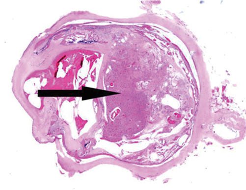 Figure 1.
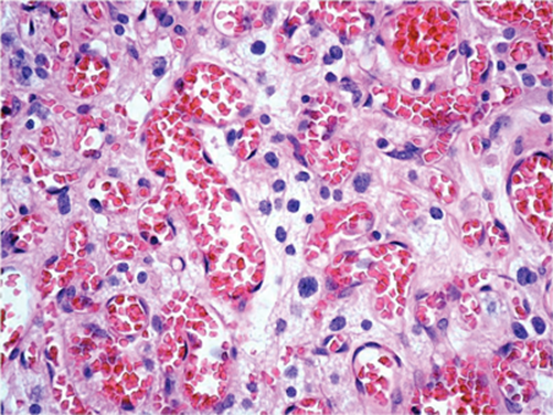
Figure 2.
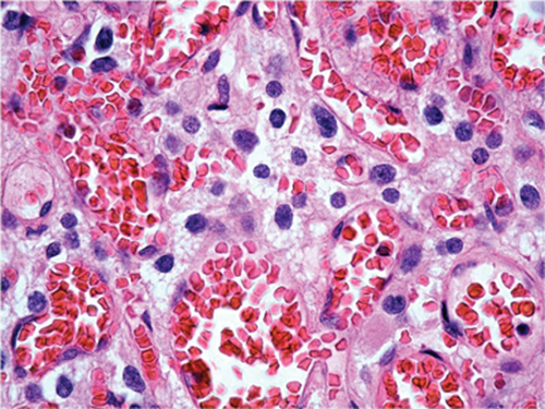
Figure 3.
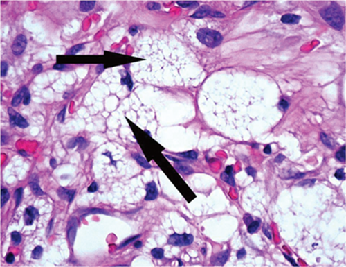
Figure 4.
Questions
-
What do Figures 2 and 3 show?
-
What does Figure 4 show (arrow)?
-
What is the likeliest diagnosis?
-
Which immuno-stain will highlight the cells in Figure 4?
-
What other history may you ask for from the clinicians?
Answers
1. A vascular neoplasm.
2. Foamy stromal cells between the vascular element.
3. The combination of endothelial and foamy stromal cells makes this a retinal haemangioblastoma. The stromal cells are the neoplastic element.
4. Anti-VEGF.
5. This patient has Von Hippel Lindau disease (hence the lesions in the other eye). The systemic manifestations are:
• Retina: haemangioblastoma
• Peripheral nerves: haemangioblastoma
• Kidney: cysts and renal cell carcinoma
• Pancreas: cysts, microcystic adenoma and neuroendocrine tumours
• Adrenal gland / paraganglia: phaeochromocytoma / paraganglioma
• Endolymphatic sac / duct: endolymphatic sac tumour
• Epididymis: epididymal cystadenoma• Adnexae: cystadenoma
• Other organs: cysts.
Further reading
- http://emedicine.medscape.com/article/1219430-overview
- WHO classification of tumours of the central nervous system. Third edition. IARC Press; 2007:215-7.
- Mettu P, Agron E, Samtani S, et al. Genotype-phenotype correlation in ocular von Hippel-Lindau (VHL) disease: The effect of missense mutation position on ocular VHL phenotype. Invest Ophthalmol Vis Sci 2010;51(9):4464-70.
COMMENTS ARE WELCOME


