Vitelliform macular dystrophies are a subset of retinal dystrophies that result in the accumulation of lipofuscin, commonly in the macula leading to the formation of characteristic vitelliform lesions [1]. Most cases have a solitary lesion in the macula, but rarely it can present with multiple extrafoveal vitelliform lesions [2].
Multifocal vitelliform dystrophy is a clinically and genetically heterogenous retinal disease that can be associated with mutations in the BEST1/VMD2 gene. It presents as bilateral multiple lesions in the fundus [3].
In optical coherence tomography (OCT) there is presence of subretinal vitelliform material and normal subfoveal choroidal thickness and vasculature. Fundus autofluorescence show hyper-autofluorescence due to the lipofuscin content in the vitelliform lesions. Genetic testing is also helpful in identifying genetic associations as the progression of disease can vary [3].
Case report
An 89-year-old male was referred to medical retina from his opticians with suspicious white lesions in his retina. This was during a routine opticians visit and the patient had no visual concerns. His prior ophthalmic history includes bilateral cataract operations. The patient’s past medical history includes epilepsy, benign prostatic hypertrophy and gastroesophageal reflux disorder. His regular medications are phenytoin, primidone and cyanocobalamin.
His vision unaided was OD 80 and OS 79 on ETDRS letters. His intraocular pressures were OD 17mmHg and OS 16mmHg. Anterior segment exam was unremarkable apart from him being pseudophakic.
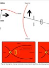
Posterior segment examination revealed mild age-related macular degeneration (AMD) in both eyes, but the more notable findings were the larger, multiple yellow-orange lesions in the superior temporal arcades in both eyes as shown in Figures 1–3.


Figures 1a (top) and 1b (above): Fundus photographs.

Figures 2.1a–2.2b: Optos FAF showed hyper-autofluorescence lesions corresponding to the fundus examination.

Figures 3a–e: OCT showed subretinal deposits corresponding with the lesions on fundus examination.
This was diagnosed as bilateral multifocal vitelliform dystrophy (MVD) and the diagnosis was explained to the patient. The patient was made aware of potential complications such as choroidal neovascularisation (CNV) and was safety-netted to seek help if his vision worsened.
Discussion
The presence of multiple yellow lesions with associated OCT findings of subretinal deposits and hyper-autofluorescence supports the diagnosis of bilateral MVD [4]. This can be considered an atypical variant of best vitelliform macular dystrophy and rarely a multifocal distribution of adult-onset foveomacular dystrophy (AFVD) [3]. Both forms have been associated with mutations in BEST1/VMD2 with only the AFVD unifocal macular subtype showing mutations in the peripherin/RDS gene [5].
Multifocal vitelliform dystrophy associated with mutations of the BEST1 are called multifocal best disease. BEST1/VMD2 codes for a bestrophin, a family of proteins, which function as a calcium dependant chloride Cl2 channel in the basolateral plasma membrane of the retinal pigment epithelium (RPE) cells. Abnormal chloride channel conductance can disrupt fluid transport across RPE and affect RPE metabolism.
This leads to accumulation of lipofuscin in and around RPE. This leads to the characteristic findings of discrete yellowish deposits in the retina with subretinal deposits in OCT and hyper-autofluorescence in fundus autofluorescence (FAF). It is important to consider other differentials such as sclerochoroidal calcification, chorioretintis, toxoplasmosis, acute exudative polymorphous vitelliform maculopathy and ocular osteoma.

Conclusion
This patient was asymptomatic and had no family history of ocular disease. He is being considered for genetic testing but extending this to his immediate family is unlikely to test for anything. There are currently no management options available, and it is important to ensure patients are followed up to monitor for complications such as CNV due to RPE atrophy.
References
1. Chawla H, Tripathy K, Vohra V. Retinal dystrophies. Florida, USA; StatPearls Publishing; 2023.
2. Querques G, Zerbib J, Santacroce R, et al. Functional and clinical data of Best vitelliform macular dystrophy patients with mutations in the BEST1 gene. Mol Vis 2009;15:2960–72.
3. Boon CJF, Klevering BJ, den Hollander AI, et al. Clinical and genetic heterogeneity in multifocal vitelliform dystrophy. Arch Ophthalmol 2007;125(8):1100–6.
4. Shah D, Saurabh K, Roy R. Multimodal imaging in multifocal best disease. Indian J Ophthalmol 2018;66(9):1313–5.
5. Crincoli E, Zhao Z, Querques G, et al. Deep learning to distinguish Best vitelliform macular dystrophy from adult onset vitelliform macular degeneration. Sci Rep 2022;12:12745.
Declaration of competing interests: None declared.