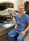
I recently had the pleasure to visit Vision Engineering’s headquarters and manufacturing facility in Woking, UK. Vision Engineering have built an international reputation for engineering microscopes that provide 3D visualisation without the use of traditional optical eyepieces.
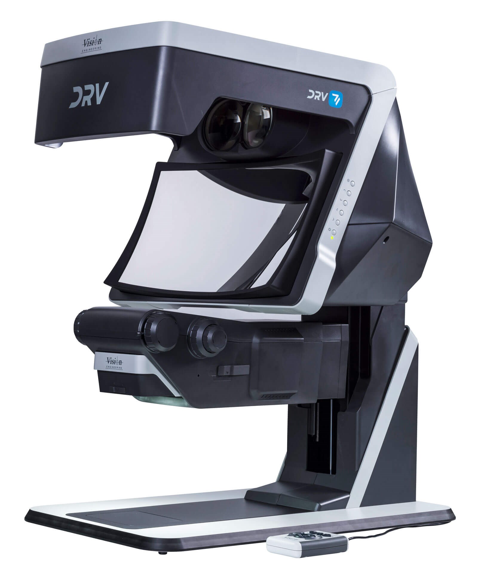
Although the company produces a range of devices using variants of this technology, the one of most relevance for this article, and ophthalmology in general, is the DRV-Z1.
The poor ergonomics and related cervical spine issues with traditional viewing systems are avoided with the Vision Engineering solutions. The technology is best tried first-hand as the view is unique. From the perspective of the examiner, the absence of eyepieces or 3D glasses / headset brings a novel science-fiction feel. Benefits observed include clinician posture, mist-free exam with facemasks, and no need to dial-in spectacle correction into the microscope, with habitual distance correction worn as needed.
The image is bright and sharp, even with high ambient lighting. There are also no artefacts, such as flickering or visible lines in the image that can be a feature of glasses-based stereo viewing systems. There is a definite head positioning sweet spot to see the image, outside of which the image is not visible. This is not a particular disadvantage, as traditional eyepieces have a very small positioning requirement.
The advantage of improved ergonomics is not overstated; I was impressed at the experience. No leaning forward or neck extension was required. Historically, this type of imaging required very high levels of illumination on the subject, making it less useful in ophthalmology. Switching from optical to digital technology has addressed this limitation, as the signal can be amplified digitally, allowing much lower levels of illumination, bringing the potential for low-light eye examinations that would be welcome by acute uveitis and similar photosensitive presentations.
This technology is now starting to gain traction within ophthalmology, with simulation leading the adoption charge. The Tennent Institute of Ophthalmology in Glasgow use the DRV as part of their wet / dry lab as an operating display.
Aside from the ergonomic advantages, these viewing systems allow accurate binocular recording. The captured video can then be replayed in the same stereo viewing system. Traditional optical eyepiece viewing and recording systems produce, at best, a reasonable monocular reproduction. The next step in the ophthalmic use of these systems will likely be in conjunction with surgical microscopes. There are many potential benefits in this area, in addition to the ergonomic improvements and the ability to record and replay accurate stereo views of the surgery. As the view is fully digital, filters, image overlays and other processing could be applied to the real-time view. Prototypes that allow binocular recording from slit-lamps are being tested and tried.
When paired with a Deep Reality Viewer they offer an excellent reproduction of the primary user’s view. With potential for digital image processing / enhancement, live digital transmission and asynchronous recorded binocular slit-lamp video streams. Further advancements include the ability to pause and rewind a slit-lamp exam when the viewing system is digitally re-presented live, which has promise for the wriggly child, capturing the fleeting glimpse of the retina or cornea. Once storage of such stereo videos is practical within ophthalmic EPR or PACS systems, clinicians would have access to accurate reproductions of historic examinations of eyes in earlier pathological states. The next step in the evolution of the slit-lamp may be in motion, potentially disrupting the present paradigm of photo-documentation.
The current versions of the DRV are large, similar to the size of a small microwave, and need to be mounted in such a way that they can be at eye level. The large size is currently a challenge, at least when considering the utility of such technology in the clinic room. If DRV technology were to replace traditional eyepieces on slit-lamps, the increased examination distance would make handheld lens holding and other interventions potentially more awkward.
Summary
Traditional technology offers monocular only surgery and slit-lamp recording. Stereo viewing systems have ergonomic issues and require glasses, headsets, or eyepieces.
The technologies described (the Deep Reality Viewer and stereo slit-lamp/surgical microscope video capture) could open a range of useful facilities to clinicians.
Surgical simulation in dry / wet labs are starting to benefit from the viewing aspect of these advances, in the form of the DRV.
The author has no proprietary or financial interests in the products discussed.
Click here for our online exlusive: Glasgow eye department continues to disrupt ocular simulation training through novel DRV system – the first of many?
COMMENTS ARE WELCOME