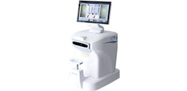
CellChek CC-20 Specular Microscope

Expand your view of the cornea with the CellChek CC-20 Specular Microscope workstation from Konan.
Easy to use – capture, analyse and report on both eyes with one click in less than 30 seconds.
Now equipped with live video of the cell layer, enabling easier capture of compromised endothelium.
Fully networkable and DICOM compatible.
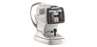
Nidek CEM-530 Automated specular microscope

CEM-530. Automated specular microscope.
The combination of central, paracentral, and peripheral imaging provides a broader view that can be used for detailed morphological and quantitative evaluation of the endothelial layer and individual cells.
- Multi area specular microscopy
- Enhanced usability and quick analysis
- Auto and manual analyses
- Additional features with optional CEM Viewer software
- 3-D auto tracking and auto shot
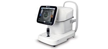
SP-1P Specular Microscope

- Wide angle ‘panorama’ photography mode: substantially increase size of the analysis area
- Two specific photography modes – sequence course and free style
- Quick automatic measurement and analysis
- Instant acquisition of the analysis result
- Intuitive operation
- Easy to read screen and comprehensive analysis software
- Frequently referred values are shown on top
- A pleomorphic / polymegethic histogram can be shown with colour
- Compact design
- 10.4” rotating touch panel monitor.
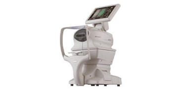
Tomey Endothelium Specular Microscope EM-4000

- Auto alignment + auto measurement
- Integrated non-contact Pachymetry
- 15 measurement areas
- Integrated database
- L-count, Trace, Core and Dark Area Method
- Counts up to 300 cells
- Integrated printer.


