Implementation of biosimilars in NHS retinal services
A NHS England (NHSE) perspective
Luke Nicholson (Moorfields Eye Hospital) explained that the NHSE perspective overall is structured on best value medicine for the patient and system, accounting for a finite budget and ensuring care for every patient/retinal disease in the system, adoption of biosimilar anti-vascular endothelial growth factor (anti-VEGF) where appropriate and effective use of longer acting anti-VEGF agents [1].
A pharmacist and medicine value perspective
Katie Joyce (North-East and North Cumbria NHS) described marked variations across medical retina services with respect to intravitreal injection treatment patterns and adoption curves for anti-VEGF biosimilars, citing capacity and scheduling challenges as well as supply disruptions. Ms Joyce emphasised the need for future proofing of treatment pathways, as best value biosimilar options may change as more biosimilar agents emerge, and there is a need to counsel patients that future biosimilar switches may be required. To capture financial savings, reinvestment conversations need to happen at the time the opportunity for efficiencies and savings is first identified.
An ophthalmologist’s perspective
Louise Downey (Leeds Teaching Hospitals) underscored the clinician’s perspective on implementation of biosimilars. The British and Éire Association of Medical Retina Specialists’ (BEAMS) position statement on biosimilar adoption provides consensus guidelines for successful implementation of NHSE commissioning guidance [2]. The consensus statement stresses that implementation must be clinically robust, allow local flexibility and allow reinvestment of savings directly into ophthalmology services.
BEAMS recommends clinicians adopt aflibercept 2mg biosimilars and ranibizumab biosimilars as first-line for neovascular age-related macular degeneration (nAMD), with phased local implementation to avoid capacity pressures. Flexibility for pressured services is recommended; where additional injections risk delays and sight loss, commissioners must permit continued use of longer-acting therapies first-line. Savings must be ring-fenced and reinvested directly into ophthalmology services. For switching policy, BEAMS recommends clinicians switch stable patients from originator aflibercept to biosimilars at the same treatment interval, with monitoring. Clinicians should not switch patients who are stable on second-line therapies. Dedicated switch teams need to be provided as support resources to manage switching large volumes of patients efficiently.
In terms of treatment intervals, clinicians should aim for extended treatment intervals of ≥12 weeks wherever clinically appropriate to minimise burden on patients and services. Clinicians should consider switching to second-generation therapies if patients fail to achieve extended intervals with biosimilars.
Repurposing older treatments for retinal disease
Which neurological and psychiatric medicines are coming soon?
Praveen Patel (Moorfields Eye Hospital) noted that repurposing medication is a valuable source of effective treatments for different medical conditions. Potential neurological and psychiatric medicines for retinal disease include L-DOPA for nAMD, tonabersat for diabetic macular oedema and fluoxetine for dry AMD.
Which metabolic and cardiovascular medicines are coming soon?
Nick Beare (Royal Liverpool University Hospital) reviewed evidence for fenofibrate for diabetic retinopathy and AMD, statins for retinal disease and elamipretide in geographic atrophy. Challenges in repurposing medicines include prospective pilot data required by funders and long study durations required for slow progressive disease. NHSE’s Medicines Repurposing Programme was suspended in April 2025, citing fewer practical repurposing opportunities than originally envisioned and few candidates possessed an evidence base strong enough to support formal license variations.
Keynote lecture: macular atrophy in dry and treated nAMD
Professor Justus Garweg (University of Bern and Swiss Eye Institute Bern) addressed macular atrophy (MA) in dry and treated nAMD in the keynote lecture. He outlined results of a retrospective, single-centre cohort study that examined potential predictors of long-term functional anti-VEGF treatment outcomes in a case series of treatment naïve nAMD patients [3]. Central retinal thickness after the anti-VEGF loading phase, time to dryness, the presence of intraretinal fluid (IRF) and the presence of MA after one year were found to predict visual function over 2–5 years.
He noted that clinical diagnosis of outer macular atrophy at baseline was not a reliable predictor of long-term functional outcomes in treated nAMD. The presence of IRF, but not subretinal fluid, after the loading phase and after one year was predictive of the visual outcome. The presence of outer MA after one year of treatment was the strongest independent predictor of visual acuity (VA) at 2–5 years, predicting vision loss of 13–20 letters over five years. Intraretinal fluid significantly predicted VA only after four years.
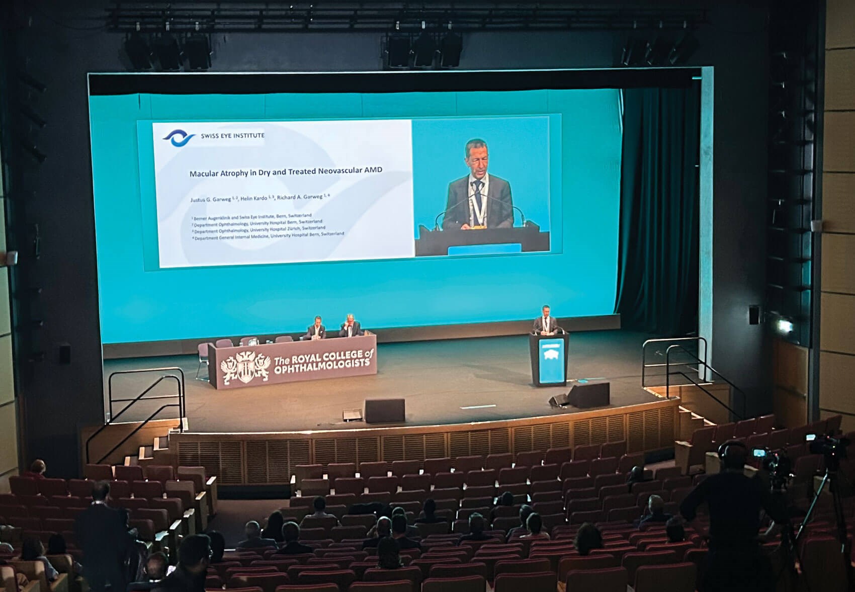
Prof Justus Garweg on the podium.
Outer retinal atrophy one year into treatment widely explains vision loss over five years, explained Prof Garweg. In this hypothesis-generating study, the progression of outer retinal atrophy was strongly linked to the presence of MA after one year and persistent disease activity, but not the number of intravitreal injections, he noted.
AI-based assessment of inner retinal layers in the same cohort revealed that the inner retinal layers, i.e. the retinal nerve fibre layer (RNFL) and ganglion and inner plexiform layers (GC-IPL) also develop neuroretinal atrophy. Although it is difficult to separate treatment effects from the underlying natural course of disease, the progression of GC-IPL atrophy was time-dependent but not associated with the number of intravitreal injections, whereas RNFL thickness remained relatively stable over up to five years. The same pattern was also observed in unaffected partner eyes.
Together, study findings suggest that AMD progression itself drives long-term loss of inner retinal layers over up to five years. Prof Garweg considered that undertreatment may be the greatest risk for atrophy and that insufficient disease control, evidenced by persistence of retinal fluid, is the most relevant predictor of long-term atrophy progression, which directly translates into vision loss.
Holistic care of diabetic retinopathy
Holistic and patient-centred management of diabetes
Sarah Jarvis (London, UK) provided an update on recommended approaches to diabetes management. Current diabetes treatment guidelines involve a combination of lifestyle modifications and pharmacological intervention. Type 1 diabetes (T1D) and type 2 diabetes (T2D) have distinct pathologies, with T2D considered as part of a spectrum of disease alongside chronic kidney disease (CKD) and cardiovascular disease (CVD), as well as associated comorbidities such as diabetic retinopathy (DR). The management of T2D requires a holistic, patient-centred approach based around glycaemic control, weight management and addressing CV risk factors. Multiple treatment guidelines recommend the use of SGLT2 inhibitors and glucagon-like peptide-1 receptor agonists (GLP-1 RAs) for the treatment of patients with T2D, CVD and CKD.
What ophthalmologists need to know about GLP-1 RA therapy
Marc Evans (Cardiff, UK) discussed results of a Delphi study designed to formulate consensus recommendations to facilitate optimum management of GLP-1 RAs in people with T2D and obesity, with a focus on those at risk of ocular complications [4]. Participants included ophthalmologists, diabetologists and obesity specialists practicing in Europe. Consensus recommendations include:
- Any new evidence on the association between GLP-1 RA or non-arteritic anterior ischaemic optic neuropathy (NAION) should be reported early
- Those at high risk of ocular events (long duration of diabetes >10 years, poor glucose control HbA1c>10%) or with established retinopathy should be screened for DR within the previous 12 months
- Any treatment intensification involving a potential rapid reduction in glucose over a short time period should be supported by retinal screening
- When prescribing GLP-1 RA to those with sight loss in one eye and or prior history of NAION, consideration of ocular risk is advised
- Clear communication between diabetologists and ophthalmologists is essential for optimum care
- Appropriate education on the natural history of early worsening of DR to increase awareness across multidisciplinary care teams is required.
The panel concluded that further research into the direct effects of GLP-1 RAs on the retina and ocular complications is required.
Vitreoretinal symposium
Management of post-intravitreal injection endophthalmitis
Felipe Dhawahir-Scala (Manchester Royal Infirmary) presented experience in the management of endophthalmitis following intravitreal injections. The ‘gold standard’ approach emphasises appropriate prevention, appropriate surgical technique, early diagnosis and prompt treatment. Current debates centre on the role of pars plana vitrectomy (PPV) and its timing for acute exogenous endophthalmitis and the use of steroids. Experience from Manchester supports immediate tap-and-inject of intravitreal antibiotics for post-intravitreal injection endophthalmitis at presentation, combined with early vitrectomy – with all patients undergoing vitrectomy within 24 hours and started on oral steroids.
Measuring success in vitreoretinal surgery
Teresa Sandinha (Liverpool University Royal Hospital) made a strong case for the development and adoption of patient-reported outcome measures (PROMS) as a core outcome of success after vitreoretinal surgery. Traditional outcome metrics of measuring surgical success rely on objective measurements of VA and anatomical features of the retina. Ms Sandinha observed that vitreoretinal surgery is highly effective at restoring vision but clinical outcomes can be unpredictable, and surgery itself carries risks and side-effects [5].
The impact of vitreoretinal surgery on quality of life is poorly studied and currently there are no validated PROMs specific to patients who have had surgery [5]. The FINESSE study group has recently developed a minimum core outcome set for idiopathic epiretinal membranes (ERMs) comprising 13 core outcomes covering six domains (measured visual function, symptoms, adverse events, functional ability, quality of life and other, including objective measurements of anatomical findings), generated by the modified Delphi process [6]. In brief, measuring success in vitreoretinal surgery should combine anatomical success, visual function, safety and validated PROMS to gauge what truly matters to patients. Future research should help define patient-meaningful change in vision and quality of life for specific vitreoretinal surgery PROMS.
Benchmarking your service: real-world evidence evaluation
The BEAVRS Database Lessons
The BEAVRS and Euretina vitreoretinal database was set up to collect rhegmatogenous retinal detachment (RRD) and macular holes outcomes for revalidation and audit. David Yorston (Gartnavel Hospital) outlined several key lessons from the BEAVRS database. In a prospective study using BEAVRS online databases of visual outcomes for macula-off retinal detachments that were successfully re-attached by vitrectomy and internal tamponade, the authors recommended recent macula-off retinal detachments should be repaired within 72 hours of the loss of central vision [7]. Surgeons should aim to re-attach the retina within four days of losing vision. The benefit of early surgery is due to better preoperative VA, reduced extent and shorter duration. Early repair increases the probability of a good visual outcome. Martins Melo, et al. reported that increasing stage of RRD was associated with worse VA at three months post-surgery [8].
On the question of choice of tamponade, Mr Yorston described findings from a study of 7466 retinal detachments treated by PPV and gas; the main outcomes were primary anatomical success and final VA. C2F6 was associated with increased odds of anatomical success compared with both SF6 and C3F8 and visual outcomes were better for both C2F6 and SF6 compared with C3F8. C3F8 was associated with a higher risk of intraocular pressure rise and higher risk of cystoid macular oedema compared with either C2F6 or SF6. Investigators cautioned that the sample is over-representative of low complexity RDs and conclusions may not apply to complex RD cases. When it comes to choosing a tamponade, surgeons should use SF6 or C2F6 rather than C3F8, concluded Mr Yorston.
National Ophthalmology Database AMD Audit
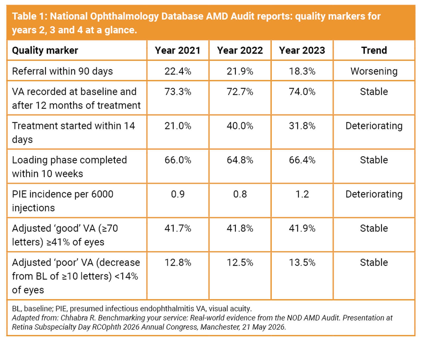
Romi Chhabra (Manchester Royal Infirmary) considered real-world evidence from the latest National Ophthalmology Database (NOD) AMD Audit, published in May 2026 (Table 1) [9]. The fourth report of the AMD audit covers patients starting treatment for nAMD in the 2023 NHS year (1 April 2023 to 31 March 2024). Results highlight significant variation in performance between different centres across the UK. The proportion of patients receiving their first treatment within two weeks of primary care referral decreased from around 40% to 31.8%, with 66.4% of eyes completing the first three injections with 10 weeks. Performance against the relevant care pathway quality markers for ‘good’ VA state after 12 months of treatment deteriorated, with 57.7% of centres achieving the acceptable level of performance compared with 67.2% in the year 2022.
Aggregate safety performance deteriorated compared with the previous year. The rates of intraocular inflammation (IOI) and presumed infectious endophthalmitis (PIE) were 2.7 cases and 1.2 cases per 6000 injections, respectively, equating to one IOI case per 2000 injections and one PIE case per 5000 injections. Performance against the PIE quality marker (<1 case per 6000 injections) deteriorated compared with the previous audit report. The proportion of centres with zero cases of PIE was 70.7%. These findings highlight the importance of robust infection prevention protocols and a structured review of all cases, notes NOD audit authors.
A total of 179,420 injections across 28,655 eyes were administered; 54.0% with Eylea (aflibercept), 29.6% with Vabysmo (faricimab), 9.7% with Ongavia (ranibizumab biosimilar), 3.0% with Lucentis (ranibizumab), 3.7% with Avastin (bevacizumab) and less than 1.0% each with Beovu (brolucizumab) and Ximluci (another ranibizumab biosimilar). For eyes starting treatment in 2023, 16.8% did not have a recorded 12-month visit, with patient death accounting for 18.4% of those absences. Loss to follow-up at month 24 was higher at 38.0%.
Recommendations from the NOD Audit report include use of a dedicated referral pathway, triage daily, start treatment quickly and ensure that initial monthly treatment is completed on time. It is recommended that clinicians use real-world data to help patients make an informed choice about starting treatment, especially when VA is poor at the start of treatment, and stop treatment when VA is <25 letters.
References
1. NHS England Biosimilars Hub. Wet Age-Related Macular Degeneration Treatment Pathway.
2. https://beams-retina.org.uk/wp-content/
uploads/2025/09/BEAMS-Position-Statement
-on-Biosimilar-Adoption-1.pdf
3. Pfister IB, Schild C, Garweg JG. Predicting long-term functional anti-VEGF treatment outcomes in neovascular AMD in a real-world setting. PLoS One 2024;19(11):e0314167.
4. Carter P, Simó R, Lövestam-Adrian M, et al. Addressing the risk of ocular complications of GLP-1RAs; a multi-disciplinary expert consensus. Diabetes Obes Metab 2025;27(12):7535–43.
5. Yoganathan A, Sandinha T, Shamdas M, et al. Patient-reported outcome measures in vitreoretinal surgery: a systematic review. Eye (Lond) 2023;37(3):391–401.
6. Sandinha T, Mehta J, Doungsong K, et al. Development of a core outcome set for patients with epiretinal membranes: a Delphi consensus study. Br J Ophthalmol 2026;110(6):651–7.
7. Yorston D, Donachie PHJ, Laidlaw DA, et al. Factors affecting visual recovery after successful repair of macula-off retinal detachments: findings from a large prospective UK cohort study. Eye (Lond) 2021;35(5):1431–9.
8. Martins Melo I, Bansal A, Naidu S, et al. Morphologic Stages of Rhegmatogenous Retinal Detachment Assessed Using Swept-Source OCT. Ophthalmol Retina 2023;7(5):398–405.
9. Gruszka-Goh MH, Monachan MT, Chhabra R. National Ophthalmology Database Audit. RCOphth 2026 [Online]:
https://nodaudit.org.uk/sites/default/files/
2026-06/NOD%20AMD%20Audit%20Full
%20Annual%20Report%202026.pdf
[All links last accessed June 2026]
Declaration of competing interests: The author has provided consultancy services to Bayer AG, DORC International B.V., Johnson & Johnson Vision Care, Inc., Roche Products Ltd and Thea Pharmaceuticals Ltd.