Whatever section of pathology is to blame and wherever it strikes, the aim of treatment is always the same.
- Find the cause if you can.
- Establish the effects of the cause.
- Halt the pathological process if you can.
- Reverse its immediate and remote effects.
- Relieve pain.
- Restore function.
The delivery of this aim follows an unchanging pattern:
- Short-term
– medical
– surgical - Long-term
– medical
– surgical
That, in essence, is the pattern of all management, simple and easy to memorise.
We are now ready to take inflammation with its old Latin tags and work out rationally what it might do to the eye, remembering that it can affect different bits of the ocular apparatus and remembering also that few conditions appear immediately in their full textbook glory. Rather, they develop with differing intensity and speed.
Rubor – redness
Calor – heat
Dolor – pain
Tumor – swelling
Loss of function – for which generally no Latin was on offer.
The process can be:
- Acute
– clearing without damage
– with replacement of special tissue with fibrous tissue. - Chronic
– Either recurrent or never clearing, with fibrosis dominant.
Applying these fundamental principles to acute iritis might go as follows:
- Red eye (Rubor)
- Hot eye (Calor)
- Painful (Dolor)
- Iris swollen: inflammatory exudate flooding anterior chamber (Tumor)
- Vision not quite right (Loss of function).
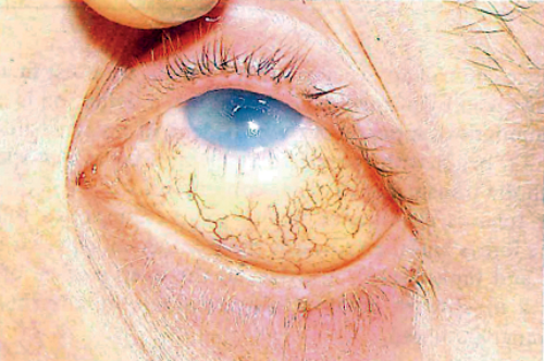
Figure 1: Ciliary injection – the major sign of a serious red eye – in this case due to keratitis,
recognised from the broken epithelium. The injection need not be florid.
All of this can be predicted from basic general pathology and each feature can be popped into its pigeon hole.
SYMPTOMS (history)
SIGNS (observations)
Now we come to the specific ocular difference – the transparent tissues:
- Exudative debris sticks to the deep corneal surface (jargon – keratic precipitates).
- A flare can be seen with a narrow beamed torch across the anterior chamber rather like dust in an old fashioned projection light.
- Aqueous flow can be disturbed in a variety of ways:
− Inflammatory exudate silting the drainage angle can block the flow of aqueous from the eye.
RESULT: Raised intraocular pressure.
− Adhesions between the iris and lens (jargon – synechiae) can block the aqueous passage into the anterior chamber. RESULT: Raised intraocular pressure.
− Prolonged disturbance of the aqueous composition willdamage dependent tissues.
RESULT: Cataract.
− If the process becomes chronic the damage continues predictably and the ciliary body could well fail in time.
RESULT: Low intraocula pressure.
We can now extend these concepts of inflammation beyond the iris and look at the behaviour and management of herpes zoster ophthalmicus in someone unfortunate enough not to have been treated in time with oral Acyclovir – a drug not yet discovered when I worked out this answer from my little system in the middle of my own FRCS examination – proof that it works under stress when lonely decisions have to be made.
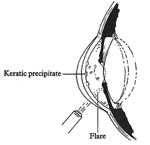
Figure 2: Inflammatory debris, flung out of solution, coats the back of the cornea and may be the only sign of iritis in a mildly inflamed eye. Such deposits are called keratic precipitates. Inflammatory exudate is not optically clear and can be picked up by a fine torch, as is dust or smoke by a projector beam.
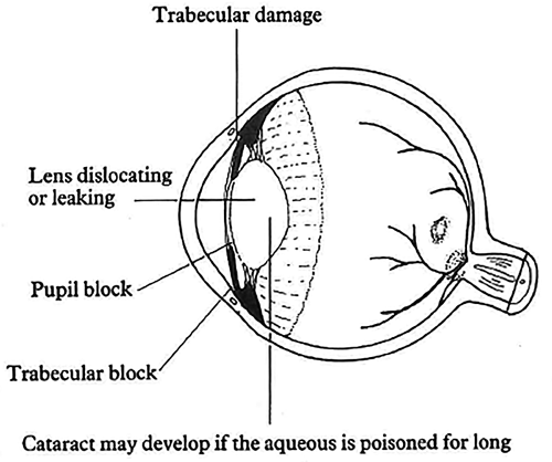
Figure 3: Acute inflammation may silt the trabecular meshwork. Chronic inflammation
may damage it with adhesions or block the pupil by the same mechanism.
Pathology
Virus induced vesicle formation along the distribution of the first division of the trigeminal nerve – thus affecting:
- skin
- conjunctiva
- cornea
- iris.
And it does not just arrive in full bloom like some textbook colour plate. It begins as vague pain, worsening along the nerve and might not be recognised until the vesicles give the game away though it might well be recognised earlier by a clinician who has come across it before. These vesicles go through all the stages of inflammation ending up as fibrosis. What happens when shingles attacks this particular nerve can now be worked out by simple reasoning. The tissues served by that nerve can be disturbed by:
- A general pathological process in a patient, likely not to be in the first flush of youth, who points you in the right direction.
- The unchanging ophthalmic ritual producing both POSITIVE and NEGATIVE findings, that will tell you − what IS wrong − what IS NOT wrong.
It is a matchlessly efficient way of using minimal information and minimal effort to create quickly a comprehensive assessment of what has happened. This is the key to the system.
Dermatitis
- Acute – all aspects of inflammation in the broken vesicles – redness, exudation and superimposed infection going on to crust formation.
- Scars developing beneath the crust. Late intractable pain.
Conjunctivitis
- Acute – pain. Discharge. Bacteria
flourishing in a viral weakened tissue.
Keratitis
- Acute. Loss of vision due to superficial punctate keratitis.
- Chronic. Corneal scars if the active lesions penetrate Bowman’s membrane. The cornea, because of its virus-induced anaesthesia loses long-term protection against incidental injury.
Iritis
- Acute. Pain. Loss of vision. Floaters. Raised pressure if the angle is silted up with inflammatory debris. NB: The patient could well have age-related undiagnosed raised pressure in both eyes, which you will pick at the sixth stopping point. Were you thinking of herpes zoster alone you might not remember this. Using the system, you could not forget.
- Chronic. Adhesions between the iris and the lens. Raised pressure, secondary to aqueous block at th pupil. Permanent damage to the trabecular network. Raised pressure.
Cataract
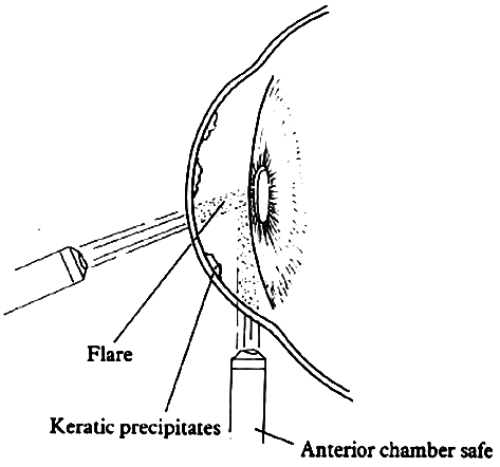
Figure 4: Iritis is the least dramatic of the serious red eyes. There is debris on the deep corneal surface and interior chamber flare. The anterior chamber is safe (eclipse test negative). Intraocular pressure may be raised if the pupil is adherent to the lens or if the drainage meshwork is clogged.
Treatment
The first step is to determine what you are dealing with and take them in order.
Dermatitis
Short-term: The aim is to kill the virus and to allow bacteria-free healing with minimal scar formation. Acyclovir skin cream five times daily deals with the first problem. Thereafter an application which combines an antibiotic with corticosteroid may achieve both these aims to some degree. Their dosage will depend on the severity of the condition.
Long-term: The pain of shingles is devastating and long-term analgesics and emotional support may still not be enough to prevent thoughts of suicide.
Conjunctivitis
The intention must be to prevent bacterial infection from spreading to the cornea with topical antibiotics in a dose related to the severity.
The cornea
Short-term: Breach of the corneal epithelium. There is no medication that can make the corneal epithelium regenerate. Conditions might be created where it would begin to recover on its own. Shingles is one of these conditions where topical corticosteroids may be permitted despite the broken epithelium. Multiple breaks in the corneal epithelium – superficial punctate keratitis – might respond if the corneal environment can be made favourable with corticosteroids. But we must remember that whilst the environment can be improved by steroids, the same drugs militate against epithelial regrowth.
“Simple memorised frameworks give you something you cannot forget in which to retrieve vital information that you cannot quite remember.”
The eye is in fact happiest when shut and closure of the eyelids with adhesive tape is the best short-term method for keeping them shut, but the eyelid skin might be too inflamed to tolerate such closure and the constant topical measures for severe iritis require the lids to be opened.
Frequent application of artificial tears goes some way to making the cornea feel that the eyelids are closed, which is its ideal state, even in health.
Iritis
The aim is to suppress inflammation and prevent complications in the acute phase. Complications and restoration of function dominate the management of the chronic phase.
Short-term: Topical corticosteroids – the dose is related to the severity of the inflammation. Mydriatics – if the pupil shows signs of adhering to the lens, then dilating it will make the development of adhesions less likely to be complete. If the intraocular pressure be raised, then it must be reduced. Acetazolomide reduces the production of aqueous. However, it also produces systemic effects in the form of increased diuresis, finger tingling, kidney stones, metabolic acidosis and possibly even mental upset.
Topical beta-blockers – these drugs also reduce the production of aqueous, but with fewer side-effects in most patients. However, they can delay the healing of corneal epithelium, and indeed may cause an intact epithelium to break down.
Long-term: The intraocular pressure may remain permanently raised and will therefore require long-term treatment, possibly with beta-blockers or other anti-glaucoma drugs, or even drainage surgery. This is the age group of chronic simple glaucoma and we must be sure that the raised pressure is indeed monocular and secondary.
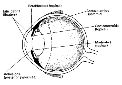
Figure 5: Correct diagnosis based on the essential symptoms and the essential signs is followed by rational treatment based on the essential principles of treatment.
Cataract
There should be no particular complication in the removal of cataract. It is always best to restore the lost focus with an intraocular lens, but if there are problems in controlling intraocular pressure before surgery, these problems may be worse after surgery, and could indeed require further surgery to control them.
Treatment before disease is established: The best management of all would be to sort all complications before they come into being. This can sometimes be achieved by systemic acyclovir if we can catch the disease in its prodromal phase. The flaw in this approach is that we must catch the condition almost before we know what it is. People tend to put up with pain in the forehead; it is only the appearance of vesicles and worse, that persuade them to seek attention. Acyclovir tablets are expensive. The dosage is four times the standard – 800mg five times daily for seven days. But treating the complications in their full flower is probably more expensive still, not to mention the price in continued misery.
A question I liked to ask in the oral examinations is how, with a torch, could you distinguish in a patient with a painful red eye with disturbed vision:
- Iritis with a secondary pressure rise from:
- Acute angle closure with secondary iritis?
The whole range of misdirected study was frequently on display, because the comparison was not described as such in any of the recommended masterworks. Slit-lamps were almost always invoked, although the question specified a torch. This is not the fault of the candidates. They are brainwashed into believing that complicated is always better and that if they remember verbatim page 800 of some textbook or other, all they need do is to share the memory with their examiner who will also know it by heart. Let us go through the ritual (see table).
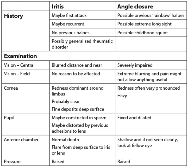
You can try the ophthalmoscope if you like but it will not tell you anything you don’t already know.
Let me give you one final story to show you how medical teaching, if dressed up with ponderous significance, can persuade students that art is indeed so long they might as well give up now. When we were having our course on surgery, I remember having one lecture taken up on a treatment for appendicitis with antibiotics. I even remember the names of the surgeons who advocated it – Ochsner and Sherrin.
It was based on the assumption that appendicitis was the result of infection rather than the cause and, of course, it didn’t work but I bet the winner of the class medal would have trundled it out to the admiration of all the examiners.
In my system it would be given five minutes as:
Short-term: medical and then would be followed pretty sharply by
Short-term: surgical.
To summarise, simple memorised frameworks give you something you cannot forget in which to retrieve vital information that you cannot quite remember.
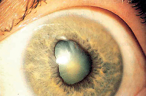
Figure 6: Acute iritis – one of the three serious red eyes. The redness is not always severe enough to be taken seriously. Inflammatory adhesions may develop between the iris and the lens, where they are given the name of posterior synechiae.
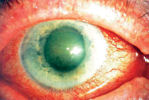
Figure 7: Acute angle closure – one of the three serious red eyes. Inflammation is dominant around the corneoscleral limbus. The pupil is fixed and dilated. The anterior chamber is shallow.

Figure 8 : Acute angle closure is one of the three serious red eyes. The cornea is hazy, the pupil fixed and dilated. The anterior chamber is shallow in each eye (eclipse test positive) and the intraocular pressure is grossly raised in the affected eye.
Acknowledgement
The images were kindly reproduced with the permission of the publisher from: Chawla HB: Ophthalmology. Churchill Livingston; 1993.
Declaration of Competing Interests: None declared.
COMMENTS ARE WELCOME