Artificial Intelligence (AI) has emerged as a prominent topic of discussion within the field of ophthalmology, captivating researchers and practitioners alike. Although recent attention has been drawn to the integration of AI in ophthalmology, it’s important to recognise that AI applications have been present in this field for several decades.

Initial applications of AI in ophthalmology primarily focussed on automating the analysis of retinal images and visual fields, paving the way for further advancements. These early endeavours set the stage for the transformative impact that AI is currently having on the discipline. In the following sections, we will explore our top five promising uses that highlight the transformative capabilities of AI in ophthalmology.
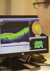
Retinal imaging analysis
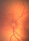
AI is being used to analyse retinal images and provide more accurate and rapid diagnoses of eye diseases. AI-powered tools can detect subtle changes in the retina and help identify disease progression at an earlier stage.
One example of this is the use of deep learning algorithms (DLA) to detect diabetic retinopathy (DR). In 2018, the Food & Drug Administration (FDA) approved the first AI-powered medical device for DR screening [1]. The device uses DLA to analyse retinal images and detect DR, which can prove particularly useful for appropriate staging and screening, with early figures of 90% specificity and sensitivity [2].
Similarly, studies such as the one conducted by Brown et al [3] have explored the application of DLA in retinopathy of prematurity diagnosis. In this particular study, a deep convolutional neural network was trained on a dataset of 5511 retinal images that were previously evaluated by experts. The results from the validation set, consisting of 100 retinal images, demonstrated promising performance. The DLA achieved a sensitivity of 93% and specificity of 94% in detecting retinopathy of prematurity (ROP) disease. Furthermore, it exhibited a sensitivity of 100% and specificity of 94% in detecting ‘pre-plus’ disease, a critical indicator of disease severity.
These findings suggest that deep learning techniques have the potential to reduce the variability and inconsistency observed in ROP diagnosis. By leveraging the power of AI, DLA can analyse retinal images objectively and provide reliable assessments of ROP disease presence and severity. Furthermore, deep learning techniques have shown promise in minimising the differences in interpretations between observers, thus enhancing diagnostic consistency and reliability.
Refractive surgery
AI-powered algorithms are being used to personalise the surgical plan for refractive surgery procedures. The AI algorithms analyse data from corneal topography, wavefront analysis and other tests to determine the optimal treatment plan for each patient, leading to better visual outcomes and fewer complications.
A key development in AI in refractive surgery is the use of femtosecond lasers. Femtosecond lasers are used to create corneal flaps in LASIK surgery, which is a type of refractive surgery [4]. AI algorithms can optimise the laser parameters, such as pulse energy and spot size, to create precise and uniform corneal flaps. AI can also determine the corneal thickness and curvature to evaluate the optimal flap size and position, reducing the risk of complications, such as corneal ectasia. Researchers have introduced various machine learning algorithms (MLA) to enhance the screening process.
Researchers have developed advanced MLA, such as the Pentacam Random Forest Index (PRFI) [5] and the ‘SCORE’ Analyzer [6], to help detect a condition called keratectasia, which can occur after refractive surgeries like LASIK. These algorithms have shown promising results in accurately identifying keratectasia by analysing data from different sources. For example, the PRFI demonstrated a sensitivity of 85.2% and specificity of 96.6% in detecting keratectasia with normal corneal shape, while the ‘SCORE’ Analyzer achieved the highest accuracy with an area under the curve of 0.911.
It’s important to note that more research is needed to fully understand the long-term effectiveness of these technologies. This will help us determine how reliable these algorithms are in predicting the development of keratectasia and ensuring patient safety after refractive surgeries.
Glaucoma screening
AI-powered tools can view images and patient data to provide remote diagnosis and treatment recommendations, allowing patients in remote areas to receive timely and accurate care. This is a key development for common pathologies, including glaucoma screening. New algorithms can identify visual field tests and retinal images to detect glaucoma and predict disease progression [7].
Researchers have explored the use of AI techniques to clarify newly developed perimeter lesions caused by glaucoma. Tarcoveanu et al. demonstrated that neural networks could discriminate primary glaucoma lesions from those caused by other diseases with an impressive accuracy of 97% [8].
It’s easy to witness this development, as in 2002, Bowd et al. ventured into the realm of AI, employing various tools to investigate the evolutionary changes in the visual field of glaucoma patients. They aimed to predict the stage of glaucoma using these AI methods. Additionally, in 2005, the same authors and contributors used two algorithms, namely relevance vector machine (RVM) and support vector machine (SVM), to classify healthy eyes from those affected by glaucoma [9]. They relied on information derived from measurements of the retinal nerve fibre layer (RNFL) and thickness obtained through scanning laser polarimetry (SLP).
These studies highlight the potential of AI algorithms in improving the diagnosis and management of glaucoma. By leveraging AI techniques, researchers and clinicians can enhance their understanding of glaucoma-related lesions, predict disease progression and distinguish glaucoma-affected eyes from healthy ones.
Drug development and use
AI is also being used to improve the efficiency and accuracy of drug development for ophthalmic conditions. AI algorithms can analyse large datasets to identify potential drug targets, predict drug efficacy and toxicity and streamline clinical trial design and recruitment.
One study used AI to analyse an anti-VEGF drug’s pharmacokinetic data and identified an optimal dosing regimen for maximising the drug efficacy and minimising the hyphenate [10], as well as predicting patient response to an anti-VEGF drug.
Furthermore, in some AI-based studies, researchers aimed to address questions related to the treatment of age-related macular degeneration, specifically determining when and if anti-VEGF therapy is necessary. Prahs et al. [11] and Schlegl et al. [12] conducted investigations using deep learning networks and found that these advanced algorithms were able to accurately predict the need for intravitreal injections in 95% of cases.
To process the large amount of data collected from baseline optical coherence tomography (OCT) scans, a DLA was employed. This algorithm utilised advanced techniques to precisely identify and analyse different parts of the OCT images, enabling it to make accurate predictions regarding the need for intravitreal injections. By leveraging the power of DLA, clinicians may be able to make more informed decisions about when to initiate anti-VEGF therapy.
Cataract management
Cataracts, a significant cause of visual impairment globally, have received relatively less attention in the field of AI compared to other age-related eye diseases. However, recent surveys have begun to explore the potential of AI in cataract detection and grading.
To detect and grade cataracts, AI techniques have been utilised with images obtained through a slit-lamp and backlit imaging, enabling the objective assessment of cataract location and severity. By leveraging DLA, precise detection and classification of cortical and nuclear changes have been successfully achieved [13].
Furthermore, AI is making significant advancements throughout the various phases of cataract surgery, including preoperative, intraoperative and postoperative stages, with a notable impact on post-surgical outcomes. AI-based modelling allows for more precise calculation of the optimal power of intraocular lenses to achieve the desired refractive outcome after surgery, surpassing traditional intraocular lens formulations in terms of precision [14]. Additionally, innovative AI-based tools for analysing surgical video footage have emerged, revolutionising the documentation, storage and cataloguing of surgical videos. These tools have found applications in teaching, training, complication review and surgical research, heralding a paradigm shift in the field.
Conclusion
From automating the analysis of retinal images to leveraging DLA for precise disease detection, the integration of AI into ophthalmology has significantly improved the efficiency and effectiveness of patient care. By harnessing the power of AI, ophthalmologists can now make more accurate diagnoses, develop tailored treatment plans and optimise outcomes for their patients. The evolving landscape of AI in ophthalmology showcases the immense potential for continued advancements in this field.
References
1. U.S. Food and Drug Administration. FDA permits the marketing of artificial intelligence-based devices to detect certain diabetes-related eye problems.
https://www.fda.gov/news-events/press-announcements/
fda-permits-marketing-artificial-intelligence-based
-device-detect certain-diabetes-related-eye
[last accessed May 2023]
2. Kumar A, Padhy S, Takkar B, Chawla R. Artificial Intelligence in diabetic retinopathy: A natural step to the future. Indian J Ophthalmol 2019;67(7):1004-9.
3. Brown JM, Campbell JP, Beers A, et al. Automated diagnosis of plus disease in retinopathy of prematurity using deep convolutional neural networks. JAMA Ophthalmol 2018;136(7):803-10.
4. Jayadev C, Shetty R. Artificial Intelligence in laser refractive surgery – potential and promise! Indian J Ophthalmol 2020;68(12):2650-1.
5. Lopes BT, Ramos IC, Salomão MQ, et al. Enhanced tomographic assessment to detect corneal ectasia based on Artificial Intelligence. Am J Ophthalmol 2018;195:223-32.
6. Chan C, Saad A, Randleman JB, et al. Analysis of cases and accuracy of 3 risk scoring systems in predicting ectasia after laser in situ keratomileusis. J Cataract Refract Surg 2018;44(8):979-92.
7. Bhartiya S. Glaucoma screening: Is ai the answer? J Curr Glaucoma Pract 2022;16(2):71-3.
8. Tarcoveanu F, Leon F, Curteanu S, et al. Classification algorithms used in predicting glaucoma progression. Healthcare 2022;10(10):1831.
9. Bowd C, Medeiros FA, Zhang Z, et al. Relevance vector machine and support vector machine classifier analysis of scanning laser polarimetry retinal nerve fiber layer measurements. Invest Ophthalmol Vis Sci 2005;46(4):1322-9.
10. Tan T-E, Wong TY, Ting DS. Artificial Intelligence for prediction of anti–VEGF treatment burden in retinal diseases: Towards precision medicine. Ophthalmol Retina 2021;5(7):601-3.
11. Prahs P, Märker D, Mayer C, Helbig H. Deep learning to support therapy decisions for intravitreal injections. Ophthalmologe 2018;115(9):722-7.
12. Schlegl T, Waldstein SM, Bogunovic H, et al. Fully automated detection and quantification of macular fluid in Oct using Deep Learning. Ophthalmology 2018;125(4):549-58.
13. Son KY, Ko J, Kim E, et al. Deep learning-based cataract detection and grading from slit-lamp and retro-illumination photographs. Ophthalmol Sci 2022;2(2):100147.
14. Lindegger DJ, Wawrzynski J, Saleh GM. Evolution and applications of artificial intelligence to cataract surgery. Ophthalmol Sci 2022;2(3):100164.
Declaration of competing interests: None declared.
COMMENTS ARE WELCOME