For trainees, having access to surgical simulation equipment is more important than ever. The authors describe the creation of a bespoke teaching and training suite in Glasgow to help trainees develop their skills and promote surgical excellence.
Simulation has become a critical component of medical training. The benefits commence from the first undergraduate clinical lessons when students learn to take a medical history and structure a clinical examination, before moving onto performing basic procedures and then with increasing complexity to the management of acutely unwell clinical scenarios. In surgical ophthalmology training, the role of simulation has been expanding dramatically over recent years [1].
Making training safer for the patient (and the trainee) echoes principles which are already enshrined in the ‘safety-first’ culture of the airline industry, where trainee and trained pilots spend many hours on purpose-built flight simulators to develop and maintain their skills. Surgical techniques (like flying manoeuvres) should also be learned and developed in a safe and supportive simulated environment, rather than on patients in the operating theatre without appropriate preparation. As technological advances have created bespoke intraocular simulation (particularly the EYESI simulator for cataract and vitreoretinal training), coupled with realistic model eyes, simulation is now an essential component of the Royal College of Ophthalmologists’ Ophthalmic Specialist Training curriculum [1,2,3].
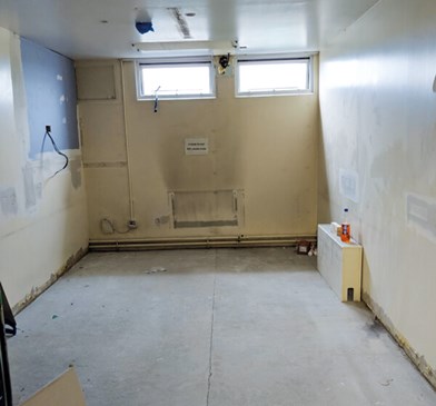
Figure 1: The original space was a long rectangle.
Renovate to innovate
Within the Tennent Institute of Ophthalmology in Glasgow, we recently had the opportunity to create a bespoke teaching and training suite. We wished to share some practical pointers to encourage other ophthalmic units to create, develop or adapt similar simulation facilities, with a view to promoting safety and excellence in training. This innovation project was funded through the NHS Greater Glasgow and Clyde Ophthalmology Endowment committee and involved renovation of a neglected library and a water-damaged computer room (Figure 1). This modernisation was designed to provide the necessary infrastructure required to promote ongoing postgraduate teaching, research, audit and surgical simulation. This was deemed to be a worthy investment in the educational requirements for the current and future ophthalmologists in Glasgow. Working with the Property and Capital Planning Department, we developed a plan to create three new rooms – a replacement computer room, a seminar library room suitable for meetings and small group teaching, and a simulation suite. Further videos discussing these developments in more detail can be found online at www.simulatedocularsurgery.com [4].
The rest of this article will focus on some practical pointers we learned from developing the simulation suite. Planning and engaging the users We wished to ensure that the facilities would be utilised by the whole ophthalmology department, so extra time walking through the renovation plans was a valuable exercise to ensure we would make best use of the space. We already had access to the EYESI simulator and an old theatre microscope, but these items did not have a permanent location and that contributed to their perceived lack of use. Additionally, there was no structure or formal provision of simulation eyes or surgical instruments for practice. We surveyed the main trainee and trainer user groups to identify what surgical simulation opportunities were lacking in the department. Through collaboration and discussion with John Ferris (RCOphth Surgical Skills Faculty Lead) and others who had experience in setting up simulation facilities or running dry labs, we were able to identify what was easily available and working well elsewhere in the UK. In particular, our trainees and trainers felt that there was a lack of time and / or equipment to practise corneal suturing and corneal incisions. Involving trainees from the beginning encouraged ownership of the facilities, which in turn can promote better engagement and use. Options for laser simulation practice were also requested, and the relevant model eyes were sourced and purchased.
We wanted a fit-for-purpose functioning simulation room which was easy to use and maintain. We decided not to bring a phacoemulsification machine into the simulation suite, and this allowed us to work in a ‘dry lab’ rather than a ‘wet lab’ environment. This was less complex to arrange around hygiene issues, and health and safety requirements, and didn’t require additional plumbing for sinks. For this reason, we would recommend that wet lab activities are reserved for the operating theatres, which are generally free for use outside of normal theatre daytime lists. In addition, we felt that the EYESI simulator already provided trainees with excellent simulated phacoemulsification training, but its ongoing use needed to be enshrined in training concepts. As the simulation room was a long rectangle shape, we split it into three areas – the surgical drylab bench, the EYESI simulator and a presentation and teaching area (Figure 2).

Figure 2: The simulation room has three distinct areas: the microscope bench;
the EYESI simulator and a presentation and teaching area.

Figure 3: Three STEMI microscopes and a repurposed theatre microscope, with variable height chairs.
Better modern equipment options
As traditional microscopes are bulky, take up a lot of space and are very expensive, we purchased three Zeiss STEMI desktop microscopes (Figure 3). These laboratory-styled microscopes are relatively affordable and mobile, yet facilitate all the hand-eye skills required for intraocular procedures. We also had previous positive experiences of these microscopes from dry labs at several different national and international ophthalmic meetings. In order to provide small group surgical teaching, we connected these microscopes to a large TV screen. We can now demonstrate surgical skills to a small group prior to supervised and solo practice (Figure 4). This TV screen is also connected to a PC and can additionally be used for surgical video presentations. We created bespoke equipment boxes relevant for individual techniques to encourage ease of use (Figure 5). As this is a dry lab with no tissue infection risks, this single use equipment can be used multiple times by many trainees. Sharps boxes are only required for used suture needles, but the provision of gloves also helps simulate the surgical environment.

Figure 4: STEMI microscopes connected to a camera for videos.

Figure 5: Bespoke equipment boxes.
Single eye, multiple procedures
We purchased a large range of the Phillips Studio eyes to supplement the microsurgical equipment [5]. We also promoted a ‘single eye, multiple procedures’ concept to maximise the use of simulated plastic eyes. This principle should help keep consumable costs down, by removing the ‘use once and throw out’ mentality. To promote this concept, we recently organised a competition to encourage trainees to be inventive, such as ‘how many ophthalmic procedures can you perform on one corneal suturing eye?’ (the current winning number is 27 and rising!). Once completed, all those sutures and / or devices can be removed and the trainee can repeat the process, all on the one model eye.

Figure 6: TV presentation screen.
Adequate storage for equipment was deemed essential to keep the area tidy and welcoming, so we installed several cupboards in the space under the workbench. We wanted to future proof the room, so provided as many electric outlets as possible with dado trunking and wireless internet connectivity. This allows presentations and videos to be shared from wireless enabled devices onto the screens (Figure 6). A new air-conditioning unit should maintain a comfortable environment for users in our Scottish winters (and slightly rarer summers). As we are working within the NHS hospital environment, we also wanted to maximise the flexibility of the room – much of the equipment is transportable should the department ever be required to move elsewhere.
At the completion of the project, we had space to place a trial lens set, so added black-out blinds to the windows to facilitate ease of refraction practice. This maximises the different uses for the room and ensures that trainees of every stage can be supported in their educational needs throughout their career.
A final important consideration is consumables. The initial financial outlay was covered by generous Endowment funding, however, ongoing purchases need planning. These ongoing purchases include plastic model eyes, sutures and surgical equipment. Through sensible planning and allocation of resources, trainees can use a set number of eyes per term. We have ongoing funding commitments from the Endowment committee who realise the cost-savings of such an approach, and a supportive training programme director who will continue to support this financially. However, it must be realised within NHS management that preventing a complication is always cheaper than the subsequent management of the same [6]. There has been a recent publication demonstrating that investing in ophthalmology cataract simulation is cost saving through the prevention of posterior capsule rupture in junior trainees [2].
Future plans
Now we have developed this simulation suite, we need to realise the benefits of this investment through regular structured use of the facility. This requires a new culture of learning to be established as part of the trainee’s commitment to learning surgical skills prior to the operating theatre, and the concept of maintaining these skills through refinement going forward. Future simulation plans include setting up individual training sessions between trainee and surgical trainer, and group teaching on specific topics (this can include anterior vitrectomy, corneal gluing, trabeculectomy, laser procedures and strabismus surgery). Providing this structure will support the Royal College of Ophthalmologists’ curriculum for simulation, and address gaps in training identified by surveys of surgical experience. Surgical logbooks only document what has been achieved by the trainee, not what may be needed, and such surveys can highlight deficiencies which can be delivered through planned simulation [7,8]. There is little value in a trainee learning how to perform a technique incorrectly, so structured supervision by senior trainees and consultants is recommended.

Figure 7: The room can be used for teaching, EYESI and microscope simulation, and refraction practise.
Our simulation suite now provides a safe environment to learn and maintain surgical skills. We expect trainees to read about surgical principles, watch online videos and then learn various techniques on the EYESI simulator before moving to the STEMI microscopes with plastic model eyes (Figure 7). They can then present their actual surgical videos in the presentation zone, where techniques can be discussed and refined in the safety of the dry lab. This stepwise development of surgical skills should ensure competence and familiarity prior to live surgery, which should also promote confidence. Trainers can also make use of the facilities to demonstrate techniques to trainees. This valuable resource should maintain a culture of constant learning and improvement and promote excellence in surgical training.
Conclusion
We hope that sharing these practical pointers will encourage other ophthalmology departments to also prioritise making ocular surgery safer, through providing modern simulation training facilities. As the suite gets used, we hope to have to purchase more STEMI microscopes to meet the increasing demand. As we slowly return to surgery from the SARS-CoV-2 enforced pause, the important role of surgical simulation is increasingly being acknowledged, and our suite is currently in almost constant use by consultants as well as trainees. At the heart of this innovation project lies a desire to strive for excellence, and to improve surgical training for the benefits of our patients. Through supportive simulation, we can teach our ophthalmology registrars basic, advanced and innovative techniques and this will modernise and enhance ophthalmology training. Promoting excellence in learning through providing the best environment to develop these skills safely should ensure the highest standards of care that our patients require and deserve.
References
1. Nairn J, Benjamin L, Lockington D. Comment on: ‘Ophthalmology Specialist Trainee Survey in the United Kingdom’. The need to increase familiarity with the management of predictable cataract surgery complications via simulation-ensuring competence to improve confidence. Eye (Lond) 2019 Nov 21 [Online ahead of print].
2. Ferris JD, Donachie PH, Johnston RL, et al. Royal College of Ophthalmologists’ National Ophthalmology Database study of cataract surgery: report 6. The impact of EyeSi virtual reality training on complications rates of cataract surgery performed by first and second year trainees. Br J Ophthalmol 2020;104(3):324-9.
3. Royal College of Ophthalmologists. Simulation in Training:
www.rcophth.ac.uk/training/
ost-information/simulation
4. Lockington D. Making Ocular Surgery Safer Through Simulation:
http://gallery.simulatedocularsurgery.com/
gallery/0/video/334
5. Phillips Studio Eye. Ophthalmic Simulated Surgery:
www.phillipsstudio.co.uk
6. Jamison A, Benjamin L, Lockington D. Quantifying the real-world cost saving from using surgical adjuncts to prevent complications during cataract surgery. Eye (Lond) 2018;32(9):1530-6.
7. Mulholland C, Lockington D. Trainee experience with capsular tension rings in Scotland-the need for structured simulation exposure to surgical adjuncts. Eye (Lond) 2020 Apr 7 [Online ahead of print].
8. Guthrie S, Goudie C, Lockington D. Survey of Scottish ophthalmic trainees’ experiences using 3-piece IOLs in cataract surgery. J Cataract Refract Surg 2018;44(3):409.
(All links last accessed June 2020)
Declaration of competing interests: None declared.
Acknowledgements:
We acknowledge the generous funding support from the Greater Glasgow and Clyde Ophthalmology Endowment committee which enabled this innovation project to come to fruition. We thank our Medical Illustration department for their help with obtaining the images (James Eyland, John Ewan) and with the supplementary videos (Adrian Struthers). We also thank John Ferris FRCOphth for publicising our work on his ocular simulation website – www.simulatedocularsurgery.com.
COMMENTS ARE WELCOME