History
-
A 73-year-old man presented with a rapidly growing left lower eyelid lump with recent bleeding.
-
On examination there was a raised, ulcerated, firm mass occupying two thirds of his lower eyelid. The lesion bled easily and appeared connected to deeper tissues.
-
Eyelid movement was restricted but no visual defects were noted.
-
Past medical history included cataract surgery and kidney transplant.
Excision of the lump was performed and specimen sent for ophthalmic histopathological assessment.
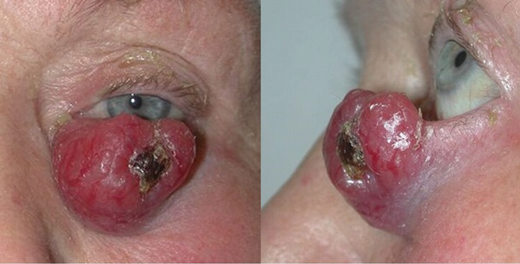
Figure 1.
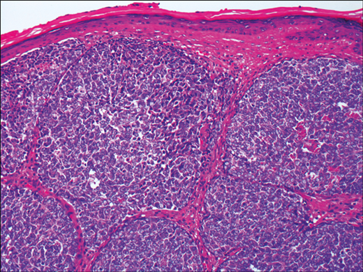
Figure 2.
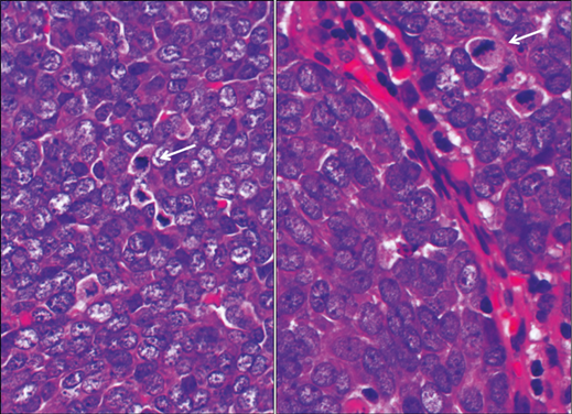
Figure 3.
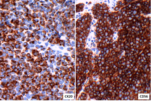
Figure 4.
Questions
-
Considering the history and clinical features (Figure 1) what is the differential diagnosis?
-
How can the histology of the excised lesion (Figures 2 and 3) be described? What do the arrows (Figure 3) show?
-
Based on features shown on Figures 2 and 3, can the differential diagnosis be narrowed further?
-
Figure 4 shows relevant markers (CK20 and CD56) expressed by tumour cells. What peculiar feature is present? Which further immunohistochemistry markers would be useful for diagnostic confirmation?
-
Taken together clinical features, histology and immunoprofile, what is the most likely diagnosis?
Answers
1. Neoplasia should always be excluded. The differential diagnosis of an eyelid tumour in an elderly patient includes carcinoma, melanoma, lymphoma and metastasis.
2. The biopsy shows a lobulated but infiltrative small cell tumour underneath stretched epidermis. Nuclei contain finely dispersed chromatin. Several mitotic figures (arrows) support high proliferation (clinical information of rapidly growing lesion).
3. The differential diagnosis of a small blue cell tumour in this age group includes lymphomas and small cell neuroendocrine carcinomas (i.e. Merkel cell carcinoma or metastatic small cell carcinoma).
4. Brown staining highlights diffuse expression of both CK20 and CD56 by tumour cells. However, the perinuclear dot-like pattern with CK20 is a useful finding seen in this tumour type. Other markers performed (not shown) to support the final diagnosis: Positive AE1/AE3 (epithelial marker), chromogranin and synaptophysin (neuroendocrine markers). Negative CK7 (epithelial marker), CD45, CD20, CD3 (lymphoid markers) and TTF1. The latter may be expressed in lung small cell carcinoma.
5. Merkel cell carcinoma.
COMMENTS ARE WELCOME


