History
-
A 67-year-old female patient had chronic left canaliculitis becoming painful and more recently complicated by left lower blepharitis.
-
Examination revealed a small fleshy lump on the medial aspect of the left lower eyelid. There was also swelling of the punctum and discharge of pus and yellow concretions upon expression.
-
Excision of the lump was performed along with canalicular curettage. Specimens sent for ophthalmic histopathological assessment.
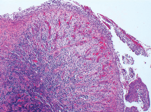
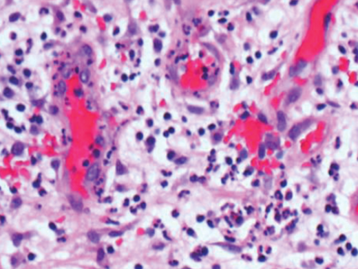
Figure 1 (top and above).
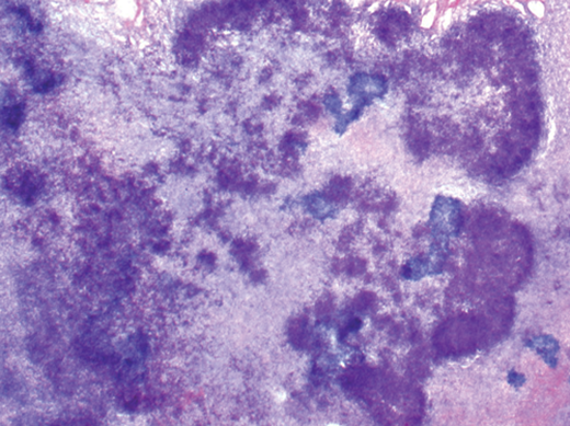
Figure 2.
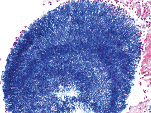
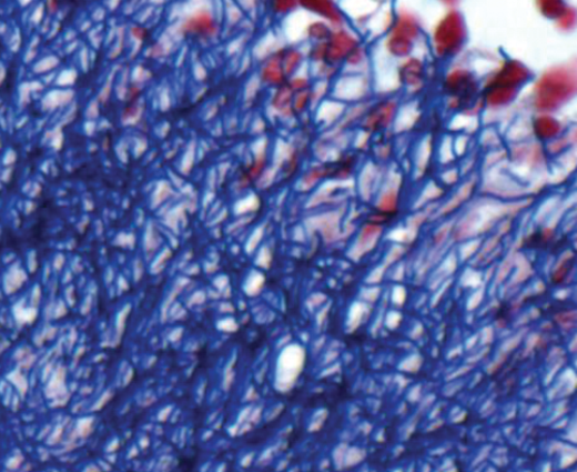
Figure 3 (top and above).
Questions
-
What does Figure 1 show?
-
Figure 2 represents H&E stained material from lacrimal sac. What is it?
-
What does Figure 3 show and which was the stain used?
-
What is the most likely diagnosis, and what is the most common causative agent?
-
Which other micro-organisms may show similar granules and how is a definitive diagnosis achieved?
Answers
1. This is a pyogenic granuloma which consists of a haemangioma-like vascular proliferation with oedematous stroma infiltrated by mixed inflammatory cells including plentiful neutrophils and usually with surface erosion.
2. The image shows amorphous hematoxiphilic material representing sulphur granules which ultimately consist of tangled filamentous bacteria. This term may be misleading, as the granules do not contain sulphur. The name actually relates to the yellow colour of the granule in pus.
3. Gram stained section demonstrating gram-positive filamentous bacteria.
4. Canaliculitis due to Actinomyces (anaerobic, gram positive, filamentous or rod bacteria). The most common causative agent is Actinomyces israelii.
5. Nocardia brasiliensis or Streptomyces madurae may show similar granules. The definitive diagnosis requires culture of the pathogen.
COMMENTS ARE WELCOME


