History
-
12-year-old cattle herder in Bangladesh.
-
Loss of vision right eye – over 2/12 to no perception of light (NPL).
-
B-scan showed choroidal thickening.
-
Suspected to be choroidal ‘tumour’.
-
Enucleated.
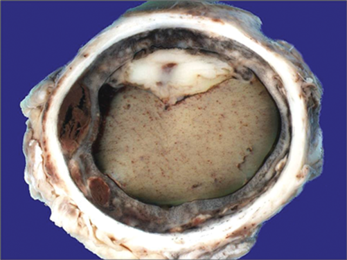
Figure 1.
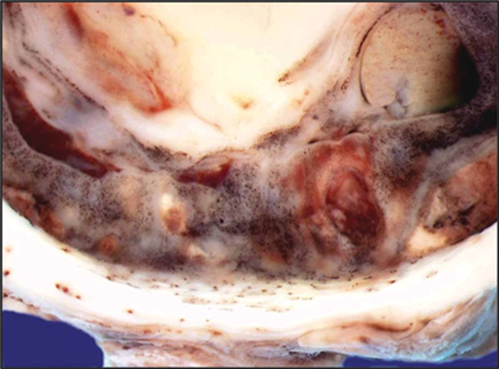
Figure 2.
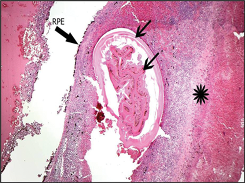
Figure 3.
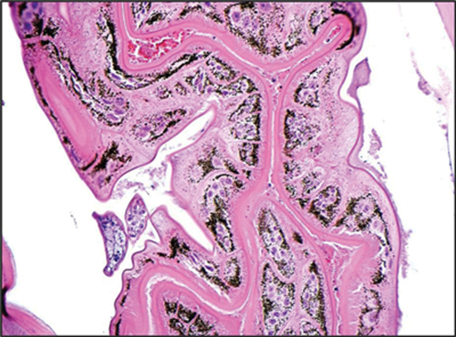
Figure 4.
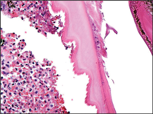
Figure 5.
Questions
-
Why is the occupation in this case of importance?
-
Figures 1 and 2 is a gross cut surface of the eyeball after formalin fixation – what does it show?
-
In histology Figure 3 – at what anatomical level is the pathology (indicated by asterisk)?
-
What is the nature of the pathology (i.e. inflammatory, infectious, neoplastic)? See Figures 4 and 5.
-
What is the likeliest diagnosis?
Answers
1. In this case, the occupation was directly relevant to the aetiology (see below).
2. It shows serpigenous, haemorrhagic tracks in the choroid and a dense vitritis.
3. The asterisk is within the choroid, which is where the pathology is. One arrow points to the retinal pigment epithelium (RPE) and the other arrows point to the pathological cause.
4. The pathology is infectious with secondary inflammation.
5. The diagnosis is ophthalmic myiasis interna. What is seen in Figures 3, 4 and 5 is a fly larva, with its internal digestive system, gonads and bosselated external layer. Figure 2 shows choroidal serpigenous haemorrhaic tracks that indicate the migration of the larva. Myiasis is the deposition of fly larvae in human tissue. Flies lay their eggs on human tissue, they hatch and the larva burrow into deeper tissues. The commonest flies to cause human ophthalmic myiasis include the latrine fly, cattle botfly, musca deomesticus (common house fly) and oestrus ovis. In our case, the child was a cattle herder and so the cause was probably the cattle botfly. The boy may have been a partial eyelid closer during sleep, allowing the flies to lay eggs on an exposed conjunctival surface.
COMMENTS ARE WELCOME


