History
-
A 35-year-old woman had a long-standing left blind eye following extensive exudative retinal detachment in the past.
-
She later developed increasing pain in her blind eye.
-
Her medical history includes pheochromocytoma resected five years ago, and she is currently on surveillance for renal cysts.
-
Her father had renal tumour and died with widespread metastatic disease.
-
Fundus examination was impaired by media opacity.
-
USB demonstrated a mass in the posterior segment.
-
Enucleation was performed.
-
Macroscopic examination revealed a partly solid partly cystic yellow-brown mass filling the posterior segment.
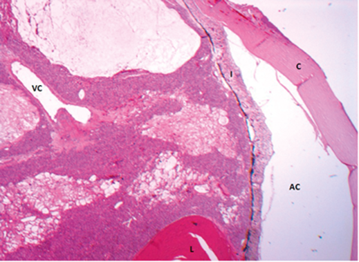
Figure 1.
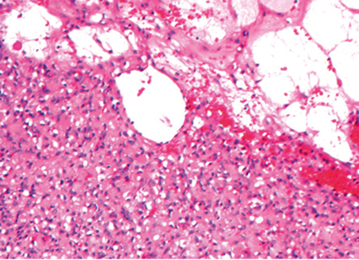
Figure 2.
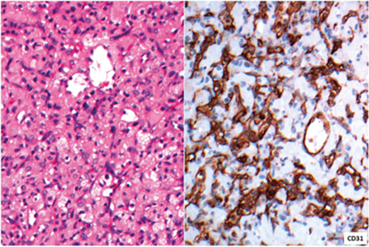
Figure 3.
Questions
-
Figures 1, 2 and 3 show representative histological and immunohistochemical sections. How can those be described?
-
What is the most likely diagnosis?
-
Considering the diagnosis on this patient’s enucleation and the clinical history provided, is there any other relevant clinical condition to be considered?
-
Are there any further diagnostic steps?
Answers
1. Figure 1 shows a solid-cystic mass filling up the vitreous cavity, encircling the lens and being adherent to the posterior surface of the iris. Figure 2: Interface between solid and ‘cystic’ component which actually represents vascular channels. Note intervening red blood cells throughout. Figure 3: Left image shows a well vascularised solid area including cells with abundant foamy and eosinophilic cytoplasm. On the right, immunohistochemistry for CD31 highlights endothelial framework in this rich vascular tumour.
2. Retinal haemangioblastoma.
3. The occurrence of retinal haemangioblastoma, pheochromocytoma and investigation for renal cysts indicates von Hippel-Lindau syndrome.
4. She most likely has a mutation on the VHL gene. The information of renal tumour in her family suggests inherited mutation. Genetic / medical screening in any sibling and children that the patient may have is essential. The patient also needs to be under close follow-up due to the risk of further neoplasia development on her only eye or other organs.
COMMENTS ARE WELCOME


