History
-
A 40-year-old male patient presented with a right lower eyelid swelling with gradual enlargement for two months.
-
On examination there was a large lesion apparently involving lid margin, part of palpebral conjunctiva and skin. No other local or systemic changes were noted.
-
An incisional biopsy was performed and the specimen sent for ophthalmic histopathological assessment.
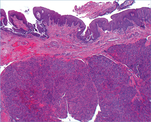
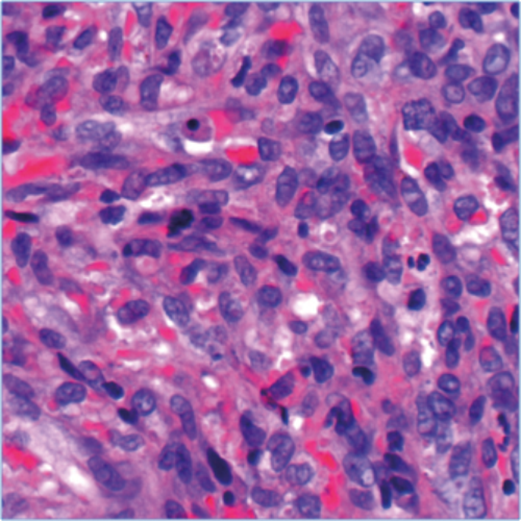
Figure 1 (top and above).
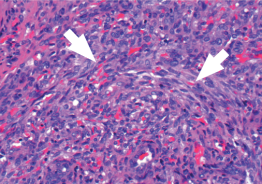
Figure 2.
Questions
-
What are the key features on the H&E (Figures 1 and 2)?
-
What do the arrows show (Figure 2)?
-
What are the initial differential diagnoses based on features seen on H&E?

Figure 3a.
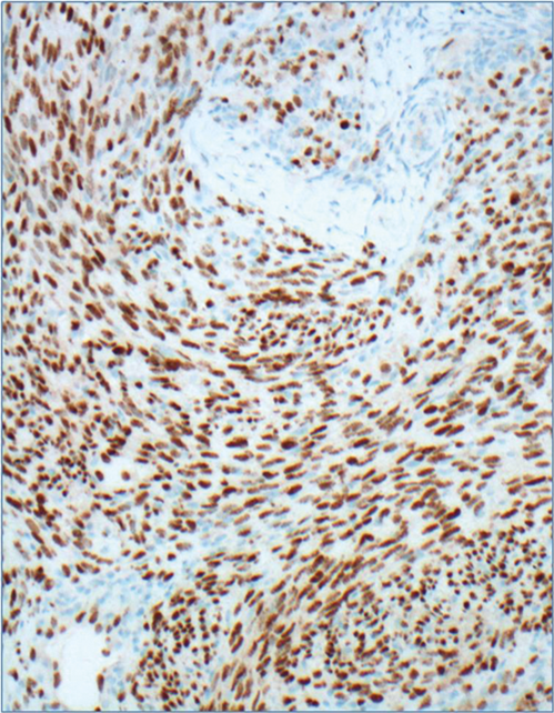
Figure 3b.
-
Tumour cells express CD34 (Figure 3a) and HHV8 (Figure 3b). Are there any other markers which could be positive in this condition?
-
What is the most likely diagnosis?
-
Which further investigations should be carried out?
Answers
1. Subepithelial dense proliferation of relatively monomorphic spindle cells. The lesion is well vascularised incorporating few slit-like vascular channels and small ill-defined blood vessels. Occasional mitoses are also seen on Figure 1 inset.
2. Extravasation of red blood cells.
3. The differential diagnosis includes bacillary angiomatosis, true vascular tumours and other spindle cell well vascularised tumours (e.g. Kaposi sarcoma, spindle cell haemangioma, Kaposiform haemangioendothelioma, solitary fibrous tumour, cellular, angiomatoid and atypical variants of fibrous histiocytoma, dermatofibrosarcoma protuberans and leiomyosarcoma). Immunohistochemistry and special stains (e.g. Warthin-Starry or gram) are required for diagnostic confirmation.
4. Other endothelial markers such as CD31, D2-40 and ERG and non-specific mesenchymal marker (i.e. vimentin).
5. Nodular Kaposi sarcoma.
6. If, like in this case, the patient was unknown to be immunosuppressed, a referral to genitourinary medicine for HIV testing and counselling would be required. Systemic investigation under the care of an oncologist is also necessary.
COMMENTS ARE WELCOME


