This site uses cookies. By continuing to browse the site you are agreeing to our use of cookies.  Read our Cookies Policy.
Close
Eye News
  • Features
    • Close
    • Features
    • Allied Professions
    • Humanitarian
    • Interviews
    • AI & Oculomics
    • Ophthalmology
    • Optometry
    • Podcast videos
    • Supplements
  • Education
    • Close
    • Education
    • Learning Curve
    • Quiz
    • Top Tips
    • Trainees
    • Medico-Legal
    • The Truth Behind The Headlines
    • Case Reports
    • Pete's Bogus Journey
  • Reviews
    • Close
    • Reviews
    • Book Reviews
    • Journal Reviews
    • What's trending?
    • Tech Reviews
    • My Top Five
    • The Culture Section
  • Events
  • News
  • Product Guide
  • Industry News
  • Contact us
    • Close
    • Contact us
    • Write for Eye News
  • Home
  • Education
  • Quiz
  • Quiz Aug/Sep 2016

Aug/Sep 2016 Quiz

1 August 2016 | EYE - Cataract, EYE - Cornea, EYE - Glaucoma, EYE - Imaging, EYE - Neuro-ophthalmology, EYE - Oculoplastic, EYE - Oncology, EYE - Orbit, EYE - Paediatrics, EYE - Pathology, EYE - Refractive, EYE - Strabismus, EYE - Vitreo-Retinal, EYE - General
Share This


History


A 45-year-old female patient presented with left upper eyelid swelling present for six months. There was mild pain and a sense of fullness over the left eye. Examination revealed a palpable firm mass over the lacrimal gland area without proptosis or associated visual impairment. An excisional lacrimal gland biopsy was performed and the specimen sent for ophthalmic histopathological assessment.


Questions


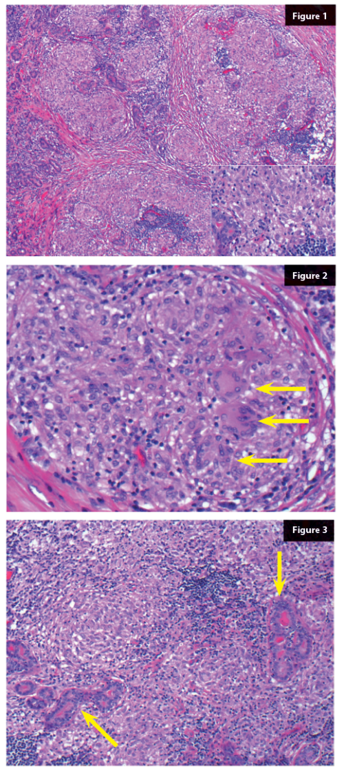
1. What are the key features on the H&E (Figure 1)?
2. What do the arrows show (Figure 2)?
3. What are the arrows pointing at (Figure 3)?
4. What is the most likely diagnosis?
5. What is the clinico-pathological differential diagnosis?
6. Which further investigations should be carried out?


Answers


1. Non-caseating epithelioid granulomas and patchy lymphocytic infiltrate.
2. Multinucleate giant cells.
3. Remainder lacrimal gland acini.
4. Sarcoidosis.
5. Other conditions with epithelioid granulomas such as tuberculosis / atypical mycobacteriosis and fungal infection.
6. Special stains for micro-organisms (Ziehl-Nielsen for AAFB and PAS / grocott for fungi). Clinical correlation is required including laboratory tests (ACE, ANCA and other inflammatory markers), radiological assessment of the chest and tuberculosis skin test.


COMMENTS ARE WELCOME

Share This
Originally Published
EYE NEWS VOLUME 23 ISSUE 2 AUGUST/SEPTEMBER 2016
Download the full article
Download Article

Top Of Page

9 Gayfield Square, 
Edinburgh EH1 3NT, UK.

Call: +44 (0)131 557 4184
www.pinpoint-scotland.com

WEBSITE DETAILS
  • Cookie Policy
  • Data Protection Notice
  • Privacy Policy
  • Terms and Conditions
ABOUT US
  • Who we are
  • Register
  • Contact us
  • Contributors
  • Company Awards
DIGITAL ISSUES/GUIDELINES
  • Digital issues - Library
  • Supplements - Library
  • Guidelines
Accreditations
IPSO_FLAG_TEAL 2025.png cpdcertified.png

Pinpoint Scotland Ltd (Registered in Scotland No. SC068684) | © 2025 - Website by Gecko Agency