History
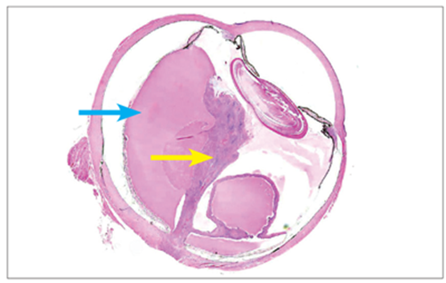
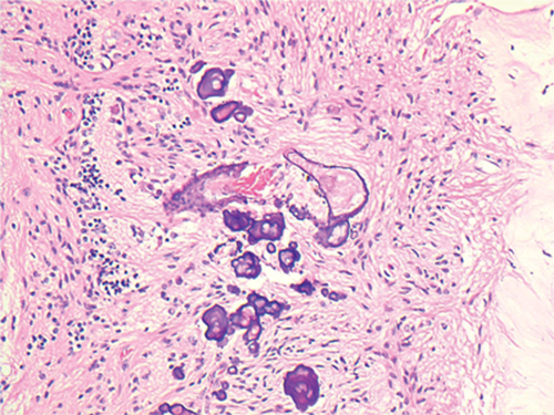
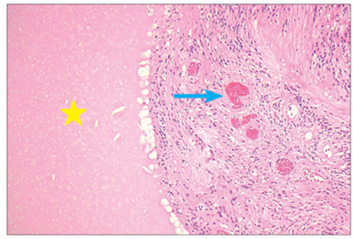
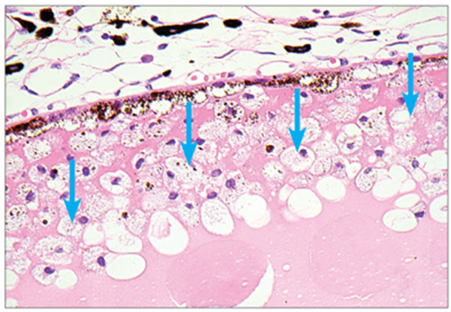
A two-year-old female child presented with loss of vision in her left eye. Examination showed leukocoria and intraocular calcification was identified on scanning. The suspected diagnosis was intraocular retinoblastoma and the child underwent an enucleation. The eyeball was submitted for ophthalmic pathology assessment. Figures 1, 2, 3 and 4 illustrate the salient histopathological features of the case.
Questions

Figure 1.
1. What are the two arrows pointing at in Figure 1?
Figure 2.
2. What is indicated by the star and arrow in Figure 2
(clue: the structures indicated by the arrow are in the peripheral, temporal outer retina)?
Figure 3.
3. What are the arrows pointing to in Figure 3?

Figure 4.
4. Which pathological process is shown in Figure 4?
5. What is the diagnosis based on the histopathological features?
6. The process of which ophthalmic conditions are present in Figure 4?
7. Are there any surprising clinical and histological features?
Answers
1. The yellow arrow is pointing to a detached retina. The blue arrow is sub-retinal serous exudate, lying between the detached retina and the sclera.
2. The yellow star is a proteinaceous serous exudate, which appears a pink colour and the arrow is pointing to abnormal vessels in the peripheral, temporal retina, containing blood.
3. These are lipid-laden macrophages near the retinal pigment epithelium (RPE). They have a frothy cytoplasmic appearance due to the high concentration of lipid material that has oozed from the vessels in the temporal retina being ingested by tissue macrophages.
4. This is dystrophic calcification of vessels. Dystrophic calcification appears as purple material deposited on the surfaces of pre-existing anatomical structures or is often seen in scar tissue.
5. Coats disease. This is based on the presence of the chronic serous exudate and the temporal, peripheral abnormal retinal vessels.
6. Retinoblastoma, retinocytoma and tuberous sclerosis.
7. The patient is a female and there is intra-retinal calcification. Coats disease affects male children usually and is characterised by the presence of abnormally dilated outer retinal vessels in the temporal peripheral retina causing chronic exudation phenomena. One of the clinical features used to distinguish Coats from retinoblastoma is the latter showing calcification. Indeed, this was the suspected indication for enucleation in this case. However, chronic Coats disease can show intra-retinal calcification. (Miller DM, Benz MS, Murray TG, Dubovy SR. Intraretinal calcification and osseous metaplasia in coats disease. Arch Ophthalmol 2004;122(11):1710-2). No viable retinoblastoma was seen in this specimen.
COMMENTS ARE WELCOME


