History
A 45-year-old man presents with left-sided epiphora. During a dacrocystorhinostomy (DCR), the wall of the lacrimal sac was noted to be bulky. The sac wall is biopsied and sent for routine histopathology examination. The pathologist notes a distinctive feature in the wall of the lacrimal sac biopsy, shown in Figures 1 and 2.
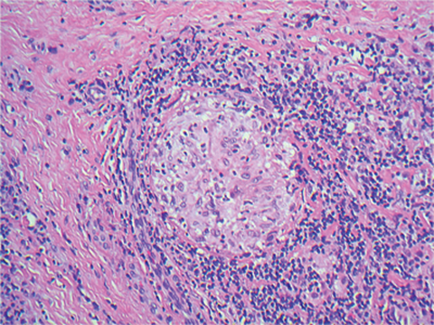
Figure 1.
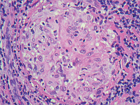
Figure 2.
Questions
1. Which stain has been used to stain the tissue in Figures 1 and 2?
2. Which cells constitute the pathology shown in Figures 1 and 2?
3. What is the name given to this type of pathology?
4. What is the differential diagnosis for this pathology?
5. What is the likeliest diagnosis?
6. Clinically, what else may you wish to enquire about?
Answers
1. Haematoxylin and eosin.
2. These are epithelioid histiocytes.
3. This is a granuloma and this is granulomatous inflammation.
4. Foreign body, infection (fungal and mycobacterial), sarcoidosis are the main differentials.
5. The patient had no infectious agents on the histology and microbiology grew no organisms. The likeliest diagnosis is sarcoidosis of the lacrimal sac.
6. Ask about systemic manifestations of sarcoidosis.
COMMENTS ARE WELCOME


