Idiopathic sclerochoroidal calcification (ISC) is a rare, benign disorder of the choroid and sclera. We present a visually asymptomatic 83-year-old caucasian male with clinical findings bilaterally of ISC, and discuss the investigations required to confirm diagnosis and help prevent further investigations which may cause unnecessary anxiety for the patient.
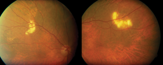
Figure 1.
Case report
ISC is a rare, benign disorder of the choroid and sclera.
An 83-year-old caucasian male was referred by his optometrist querying the cause of bilateral superotemporal fundal lesions. The patient was visually asymptomatic with an unremarkable past medical history. Clinically visual acuity was Right: 0.36 logMAR improving to 0.26 logMAR with pinhole and Left: 0.6 logMAR improving to 0.04 logMAR with pinhole. The anterior segment assessment showed moderate cortical lens opacity bilaterally and dilated fundoscopy revealed evidence of slight raised yellowish lesions (Figure 1) on the superior aspect of the superotemporal arcade bilaterally. B-scan ultrasound examination revealed highly reflective lesions and confirmed the clinical diagnosis of bilateral sclerochoroidal calcification (Figure 2).
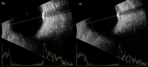
Figure 2.
Discussion
Deposition of calcium in this nature was initially described by Pagenstecher in 1860 [1]. Its clinical differential diagnosis includes choroidal osteoma, choroidal metastasis, choroidal melanoma, choroidal amelanotic naevus and choroiditis [2]. The lesions are mainly observed in the mild-peripheral fundus close to the superotemporal arcade as multiple, well-defined, elevated yellow plaque-like lesions [3]. The lesions have echographic patterns consistent with calcification which are often documented in both the choroid and sclera. Lesions have frequently been documented in the choroid alone but never alone in the sclera [4]. It is important to highlight that patients who have such lesions also have normal calcium metabolism.
Sclerochoroidal calcification is a very rare but well recognised ocular condition that is believed to be idiopathic in most cases and unlikely to cause symptoms or visual problems to the patients. Its incidence is higher in the caucasian population [5]. Usually seen in elderly patients otherwise visually asymptomatic, ISC is often an incidental finding and the lesions are typically bilateral and multifocal. They can have either plaque-like or tumour like appearance (sometimes up to 6mm in height) hence the importance of early diagnosis [6]. Fundal examination and the specific echography pattern is usually sufficient to arrive at a clinical diagnosis with fluorescein angiography being a useful tool in atypical lesions. This said, if misdiagnosed, this condition may lead to extensive and unnecessary further investigations [7].
References
1. Watson PG, Hazleman BL, McCluskey P, Pavésio CE. The sclera and systemic disorders, 1st edition. JP Medical;2012:276-7.
2. Sivalingam A, Shields CL, Shields JA, et al. Idiopathic sclerochoroidal calcification. Ophthalmology 1991;98(5):720-4.
3. Pakrou N, Craig JE. Idiopathic sclerochoroidal calcification in a 79-year-old woman. Clin Experiment Ophthalmol 2006;34(1):76-8.
4. Schachat AP, Robertson DM, Mieler WF, et al. Sclerochoroidal calcification. Wilmer Ophthalmological Institute, Baltimore, Md Arch Ophthalmol 1992;110(2):196-9.
5. Honavar SG, Shields CL, Demirci H, et al. Sclerochoroidal calcification. Arch Ophthalmol 2001;119:833-40.
6. Leys A, Stalmans P, Blanckaert J. Sclerochoroidal calcification with choroidal neovascularization. Arch Ophthalmol 2000;118:854-7.
7. Lim JI, Goldberg MF. Idiopathic sclerochoroidal calcification. Arch Ophthalmol 1989;107:1122-3.
COMMENTS ARE WELCOME


