
Epiphora secondary to acquired nasolacrimal duct obstruction (NLDO) is a common presentation in ophthalmic practice, with an incidence of 30.47 per 100,000 population [1]. Histopathological studies demonstrate that NLDO is characterised by chronic inflammation and progressive fibrosis of the ductal mucosa, resulting in partial or complete luminal obstruction [2].
Lacrimal syringing plays a key role in localising the level of obstruction. A ‘hard stop’ during syringing occurs when the cannula tip abuts the lacrimal bone, indicating patent canaliculi with distal obstruction at the lacrimal sac or nasolacrimal duct. In contrast, a ‘soft stop’ suggests canalicular or proximal pathology.
Endonasal dacryocystorhinostomy (DCR) is an established technique creating a direct anastomosis between the lacrimal sac and nasal cavity. This report describes a technical manoeuvre using viscoelastic injection to facilitate intraoperative identification and controlled distension of the lacrimal sac, enabling precise medial wall incision while minimising trauma and postoperative scarring.
Case report
A patient presented with persistent watery and mucopurulent discharge from the left eye. Lacrimal syringing demonstrated a ‘hard stop’, indicating distal obstruction. A joint endonasal DCR was performed by the ear, nose and throat (ENT) and ophthalmology teams.
Nasal endoscopy using a rigid 0-degree endoscope was undertaken following nasal decongestion and topical vasoconstriction. An incision was made in the lateral nasal mucosa overlying the lacrimal fossa, and the lacrimal bone with part of the frontal process of the maxilla was removed to expose the medial wall of the lacrimal sac.
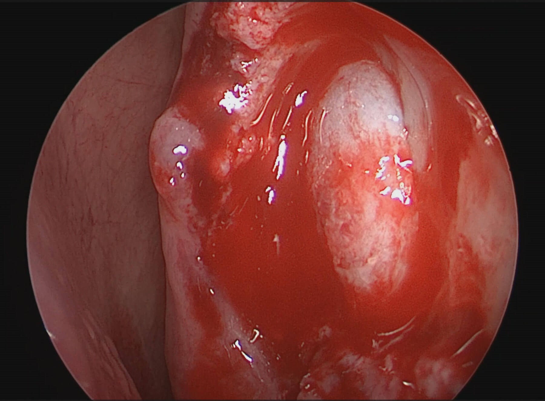
Figure 1: Intraoperative nasal endoscopic image demonstrating distension of the nasolacrimal sac with viscoelastic, producing a smooth bulge in the lateral nasal wall.
A viscoelastic agent was injected via the lacrimal punctum, distending the nasolacrimal sac and producing a tense, well-defined bulge visible endonasally (Figure 1), confirming its location and facilitating identification.
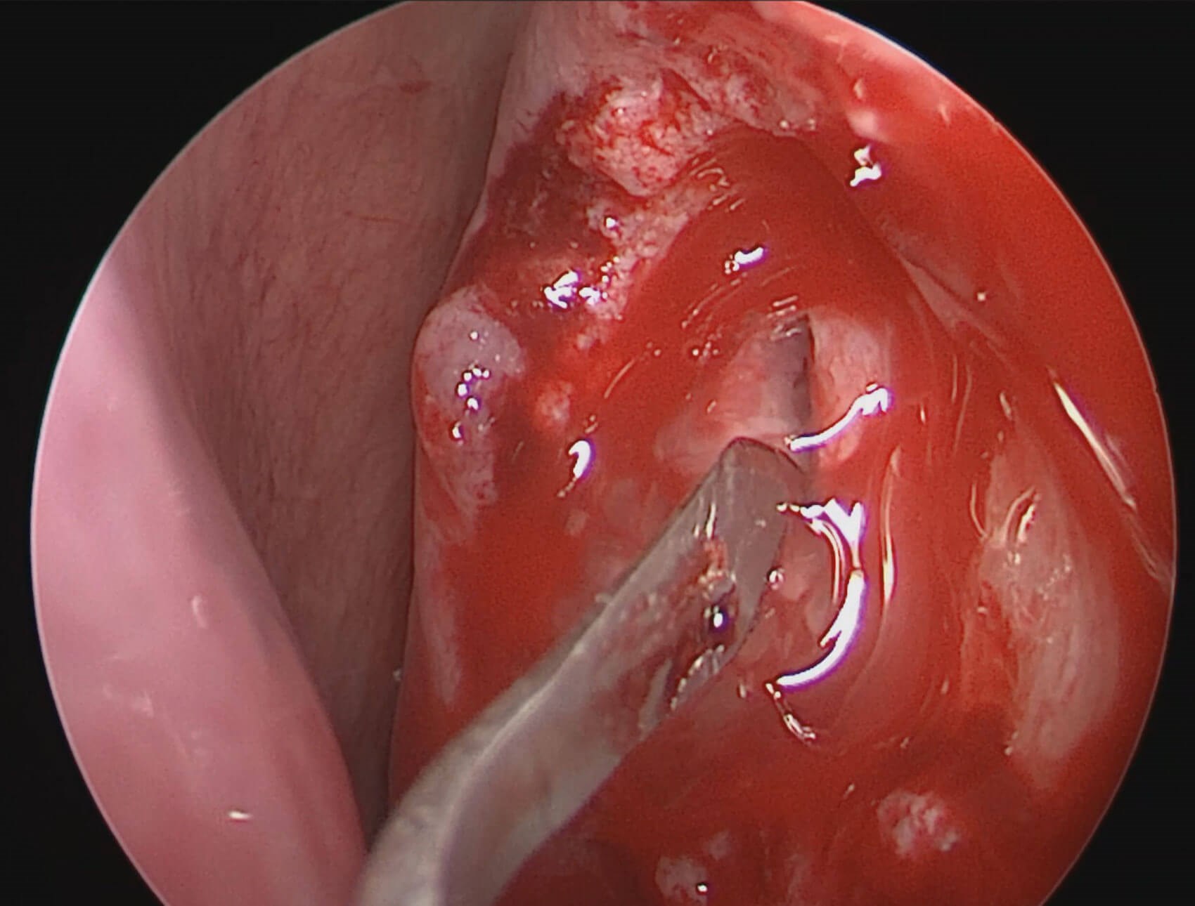
Figure 2: Nasal endoscopic photograph showing the keratome blade incising the anterior wall of the inflated nasolacrimal sac, with release of viscoelastic material.
The medial wall was then incised using a keratome under endoscopic guidance (Figure 2). Release of viscoelastic gel into the nasal cavity confirmed successful entry into the sac lumen. A bicanalicular silicone stent was inserted and retrieved intranasally to maintain patency of the ostium.
The procedure was uncomplicated. Postoperative care included intranasal corticosteroids and saline irrigation, with planned stent removal at 6–8 weeks. Postoperative stent removal was uneventful, and the patient had a successful outcome during short-term follow-up.
Discussion
Dacryocystorhinostomy bypasses NDO by forming a direct communication between the lacrimal sac and nasal cavity. The endonasal approach avoids a cutaneous scar and allows direct visualisation, with reported success rates comparable to external DCR (approximately 75%) in selected patients [3].
A joint ENT-ophthalmology approach is commonly adopted: ENT surgeons contribute expertise in endoscopy and rhinostomy formation, while ophthalmologists perform lacrimal cannulation, stenting and anatomical assessment [4].
Ophthalmic viscosurgical devices (OVDs), introduced in 1972, are widely used in intraocular surgery due to their viscoelastic properties [5]. Cohesive agents, such as sodium hyaluronate (e.g. Healon®), have high molecular weight and maintain space under pressure, making them well suited to distending enclosed compartments. Dispersive agents, such as hydroxypropyl methylcellulose (e.g. OcuCoat®), have lower viscosity, adhere to tissue surfaces and are more resistant to removal.
We used sodium hyaluronate to distend the nasolacrimal sac, producing a smooth, tense bulge within the lateral nasal wall, aiding identification and controlled incision with reduced tissue handling and minimising inadvertent injury to adjacent structures, including the lateral sac wall, which may otherwise predispose to scarring and occlusion.
Limitations include the additional cost of OVDs and potential lack of availability in ENT settings, requiring cross-specialty coordination. Furthermore, this technique represents local innovation, with no published comparative studies evaluating outcomes with and without viscoelastic use. Future case series and comparative studies are warranted to assess intraoperative safety and long-term patency outcomes.
In conclusion, this case highlights the importance of accurate lacrimal syringing in evaluating epiphora and the need for precise intraoperative localisation during DCR. Viscoelastic distension of the lacrimal sac provides a simple adjunct to improve identification, facilitate controlled incision, and reduce tissue trauma. Further evaluation is required to determine its impact on surgical outcomes.
References
1. Woog JJ. The incidence of symptomatic acquired lacrimal outflow obstruction among residents of Olmsted County, Minnesota, 1976–2000 (an American Ophthalmological Society thesis). Trans Am Ophthalmol Soc 2007;105:649–66.
2. Linberg JV, McCormick SA. Primary acquired nasolacrimal duct obstruction: a clinicopathologic report and biopsy technique. Ophthalmology 1986;93(8):1055–63.
3. Hartikainen J, Antila J, Varpula M, et al. Prospective randomized comparison of endonasal endoscopic dacryocystorhinostomy and external dacryocystorhinostomy. Laryngoscope 1998;108(12):1861–6.
4. Wormald PJ. Powered endoscopic dacryocystorhinostomy. Laryngoscope 2002;112(1):69–72.
5. Liesegang TJ. Viscoelastic substances in ophthalmology. Surv Ophthalmol 1990;34(4):268–93.
Declaration of competing interests: None declared.





