In today’s world, macular laser treatment has a vital role in the treatment of diabetic macular oedema (DMO). DMO is one of the most common causes of visual impairment. Despite expensive intravitreal treatment courses of anti-VEGF, many will agree that focal and grid macular laser treatments have been a gold standard of care; essential for treating oedema and retinal thickening [1].
The main rationale for laser treatment is to seal leaking blood vessels and also to reduce and stop new vessels from developing. Therefore, the latter is achieved using full panretinal photocoagulation (PRP) and the former with focal laser treatment, which is commonly used when administering macular laser treatment. This article aims to provide helpful and practical tips when applying both macular and PRP laser treatment for the treatment of diabetic retinopathy and maculopathy.
Factors to be considered include patient positioning and comfort, preparation prior to treatment, slit-lamp examination findings and considerations, laser settings and type, technique used for delivering treatment and ensuring a low complication rate.
Patient position and comfort
This is essential to ensure a successful treatment session. Patients must be comfortable in the first instance [2]. This will mean that patients will be able to concentrate and adhere to instructions given by the clinician.
Patient position can be optomised by ensuring that patients are sat at the right position on the slit-lamp. A few extra minutes to get this stage correct will prove invaluable. The patient should be at the right height to make sure that there is no cramping of the neck or tilting of the patient’s head. This will also help ensure that their forehead is touching the bar throughout the treatment session.
Pain is often one of the largest fear factors – particularly with PRP laser and potentially long treatment sessions – which patients have, resulting in anxious, worried and restless patients. As a result, the treatment session takes longer and is less successful. One of the reasons for pain is thought to originate from photocoagulation of the ciliary nerves traversing in the supra choroidal space [3].
To maximise comfort, anaesthesia must be applied at an adequate level and escalated as necessary. Every patient should have a topical anaesthetic, e.g. proxymetacaine or tetracaine. A study by Al-Hussainy et al. [4], recommended treatment to the fellow eye also as this would reduce ocular surface irritation and dryness in the fellow eye resulting from prolonged treatment sessions. They explain that this may also reduce photophobia, which may develop secondary to holding the eyelids open to apply the treatment. Furthermore, this study demonstrated that the application of topical anaesthetic to both eyes aids the fellow eye in holding a fixed position of gaze and reduces Bell’s phenomenon. This is extremely beneficial when treating a particular section of the retina and provides an added safety measure as patients can be instructed not to look at the laser dot.
Taking oral paracetamol, 1g two hours before laser therapy, may be effective in the majority of patients in reducing the pain from PRP after the treatment has been given [5].
Topical anaesthesia alone may not be sufficient in all patients and therefore adjunct more invasive anaesthetic may be required; these include a sub tenon [6] block and very rarely a retrobulbar or peribulbar block is administered.
Preparation
Having attended to the patient and ensured that they are comfortable and positioned correctly, one must ensure that all the equipment to be used is also optimal. Successful laser treatment is highly dependent on accurate placement of the laser on the retina. This can only be achieved with a stereoscopic view.
Therefore, it is vital to adjust the eyepieces on the slit-lamp to fit the operators’ interpupillary distance; to ensure a sterioscopic view of the retina for the entire duration of your laser therapy session. This can also be made more accurate by using the focusing rod to fine tune the focusing prior to starting treatment.
Examination findings and considerations
Examination of patients prior to beginning treatment is essential as this will determine various settings and parameters for laser treatment. Examine the patient’s anterior segment specifically to identify pathology such as: corneal scaring, oedema, lens opacities, pupil size and posterior synechiae. In turn, the laser settings and parameters used will ultimately have a large bearing on the success of your laser therapy. Furthermore, in cases of media opacities (of any cause) one should resort to higher powers to achieve adequate laser burns.

Figure 1: Common Volk contact lenses used for administering retinal laser treatments [7].
The most common contact slit-lamp lenses (Figure 1) used to administer retinal laser treatment in most ophthalmology departments are listed in Table 1. Each lens has its own merits and indications.

When performing laser treatment over a period of 10 minutes it is not unusual for the view to be hazy and blurred. This is due to escape of the coupling fluid beneath the contact lens. We advise that reapplication of the contact lens with fresh coupling gel will greatly improve ones’ view. If the view of the retina at the time of applying the laser spot is out of focus or lost then the background illumination of the slit-lamp beam should be increased and / or widened. One’s view may also be impaired due to corneal decompensation secondary to rubeosis and raised intraocular pressure with corneal oedema.
The location of required laser treatment is also paramount to successful treatment. As central retina is thicker than peripheral retina; as one moves from the centre to the periphery the power should be reduced to avoid any severe laser burns. It is pertinent that there is good PRP coverage far out in the peripheral retina. This can be achieved with the aid of wide-field optos images when planning treatment. However, where this is difficult or not possible, indirect laser treatment may be indicated.
There are a wide variety of complications that may be caused by severe laser burns such as choroidal neovascular membranes, larger scars, laser scar extensions due to confluence, visual field defects and increased retinal ischaemia. In certain scenarios, confluent laser treatment may be deliberately sought during PRP. We recommend confluent laser therapy to the areas occupied by neovascular fronds as long as they are well away from the posterior pole. This will help to resolve new vessels elsewhere, completely preventing further vitreous haemorrages and formation of vaso-proliferative masses.
Age is all important
It is a well-known fact across a wide range of specialties that younger patients may be more apprehensive and more pain sensitive. This is also true in the world of ophthalmology and must be taken into account when applying retinal laser treatment. These patients require less laser power to produce effective laser burns on their retina. This is mainly because of the fact that their crystalline lenses will transmit the majority of the laser energy, in contrast to an older person with lenticular sclerosis. Therefore, reducing the laser power for a younger patient and increasing it for an older patient will achieve similar, adequate laser burns on the retina.
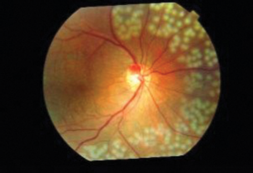
Figure 2: Retinal laser treatment, indicating the spacing between laser burns [8].
Technique – mind the gap and shape
The spacing between retinal laser burns is important (Figure 2); particularly in the case of macular laser treatments; with half to one laser spot size spacing between shots. Confluent laser burns produce permanent central visual field defects and this can have a significant impact on patients, for the rest of their life, on a daily basis. Minimising this is therefore vital, not only to ensure that the patient’s quality of life is unaltered, but also to aid with adherence to treatment and encourage patients to attend further appointments, some of which may be for further laser treatment. We all should consider the prospect of enlargement and stretching of the laser burns with age and time, at the time of applying these essential treatments; as this factor alone can cause loss of vision at a later date in the patient’s lifetime.
In general, the laser spot should be round in shape at the time of application to achieve an effective laser burn. When the spot is round, all the intended laser energy is transmitted to the correct spot in the fundus. However, if the laser spot is oval, crescent or any other shape then part of the laser energy is lost in the media and it is difficult to produce an effective laser burn on the retina. Odd shaped laser spots normally denote loss of coaxial view of the fundus due to excessive and abnormal tilting of the lens on the eye. This is the case with both macular and PRP laser treatments.
Laser settings and type
Indirect laser treatment has specific indications and can result in better views of the fundus. There are various techniques which one can employ to maximise view, such as, a 28 dioptre lens when patients have a small pupil; glycerin is extremely useful in reducing corneal oedema. This method of laser treatment application also provides added stabilisation of the globe if a scleral depressor is used. In comparison the slit-lamp allows for increased precision and placement of laser burns, enabling the operator to control the amount of space in-between laser shots.

Laser treatment settings will always be variable and based upon the clinical situation, indication and patient factors. However, there are certain basic guidelines, which provide a framework for treatment settings. These are summarised in Table 2 from Diabetic Retinopathy Study (DRS) and Early Treatment Diabetic Retinopathy Study (ETDRS) studies. It is proposed that this treatment is applied over two sessions [9]. Furthermore, specific ischaemic portions of retina maybe treated based on fundus fluorescien angiography (FFA) results.
“Given the current era which is heavily biased towards anti-VEGF treatment, it is vital to ensure that appropriate measures are taken to optomise and perfect retinal laser treatment for diabetic patients.”
Macular grid laser essentially refers to multiple laser shots applied to the macula, sparing the fovea, which are widely spaced. Modified macular grid laser aims to try and specifically target areas of microaneurysms and oedema. Furthermore, the correct laser spot size is another critical factor for successful treatment. For focal macular laser 100 microns is an adequate size, whereas for grid laser therapy one can use between 100 and 200 microns.
More recently macular laser treatment has been performed using a modified laser technique which includes modified ETDRS and more extensively modified macular grid laser. A study comparing both techniques in 2015 demonstrated that, at one year, the modified macular grid technique was less effective at reducing retinal thickening (from optical coherence tomography (OCT) findings). Concluding that a longer-term trial examining this technique was not justified [10].
For panretinal laser therapy, spots over 300 microns would help to cover wider areas quickly. A shorter duration is advised if the patient is experiencing any pain or discomfort or moving excessively. This is less painful and in turn would enable the operator to maintain the same power settings. A linear laser spot instead of a round spot will result if there is any eye movement during the application. This must be taken into consideration when applying loser spots close to the foveal avascular zone. If the patient blinks or squeezes the eye during treatment then the eye rolls up due to Bell’s phenomenon and the laser spot may inadvertently extend and be applied to the fovea with catastrophic consequences. Therefore, we advise that peripheral retinal laser is initially applied to the upper half until certain that there are no untoward or unexpected eye movements.
Conclusion
Given the current era which is heavily biased towards anti-VEGF treatment, it is vital to ensure that appropriate measures are taken to optomise and perfect retinal laser treatment for diabetic patients. These various tips, tricks and pearls of knowledge from a wealth of experience will help achieve this and, if not reduce the need for anti-VEGF treatment, will be a successful adjunct to this more invasive treatment.
References:
1. Dowler JF. Laser management of diabetic retinopathy. J R Soc Med 2003;96(6):277-9.
2. NICE guidelines [CG138]. Patient experience in adult NHS services: improving the experience of care for people using adult NHS services. NICE. 2012.
3. Bloom SM, Brucker AJ: Laser Surgery of the Posterior Segment. 2nd ed. Lippincott-Raven: Philadelphia; 1997.
4. Al-Hussainy S, Dodson PM, Gibson JM. Pain response and follow-up of patients undergoing panretinal laser photocoagulation with reduced exposure times. Eye (Lond) 2008;22:96-9.
5. Vaideanu D, Taylor P, McAndrew P, et al. Double masked randomised controlled trial to assess the effectiveness of paracetamol in reducing pain in panretinal photocoagulation. Brit J Ophthalmol 2006;90(6):713-7.
6. Stevens JD, Foss AJ, Hamilton AM. No-needle one-quadrant sub-tenon anaesthesia for panretinal photocoagulation. Eye (Lond) 1993;7:768-71.
7. Volk Training Presentation.
http://www.slideshare.net/kevainen/
volk-training-presentation-ns
Last accessed April 2017.
8. Sundar Eye Hospital Photo Album of Diabetic Retinopathy Patients.
http://www.sundareyehospital.org/
photoalbumofourdiabeticretinopathypatients.html
Last accessed April 2017.
9. Early Treatment of Diabetic Retinopathy Study Group. Early Photocoagulation Study Group. Techniques for scatter and local photocoagulation treatment of diabetic retinopathy: the Early Treatment of Diabetic Retinopathy Study report no. 3. Int Ophthalmol Clin 1987;27:254-64.
10. Diabetic Retinopathy Clinical Research Network. Comparison of Modified-ETDRS and Mild Macular Grid Laser Photocoagulation Strategies for Diabetic Macular Edema. Arch Ophthalmol 2007;125(4):469-80.
COMMENTS ARE WELCOME


