Optos, Plc, the leading retinal imaging company, announces it is expanding the optomap® ultra-widefield (UWF™) retinal imaging modalities available with the California FA device to further assist eyecare professionals in disease management and treatment planning.
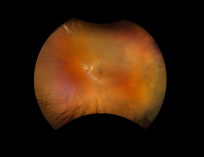
In addition to optomap colour rg (red/green), sensory red-free, choroidal, autofluorescence (AF), fluorescein angiography (FAF) and indocyanine green angiography (ICGA) modalities, the company is pleased to announce the first ultra-widefield colour rgb (red/green/blue colour) image. This new image modality is captured simultaneous to the optomap colour rg image. Thus, a single capture delivers two amazing colour ultra-widefield images.
Rob Kennedy, CEO of Optos, said: “We are pleased to bring another image modality to our California FA device. The optimised colour palette used in the new optomap ultra-widefield colour rgb image was created in close collaboration with eyecare professionals. The new image modality, in addition to our clinically validated optomap colour rg images, provides additional retinal visualisation to our customers as they manage their patient’s treatment and ongoing care.”
Initial feedback from customers who saw the new image at the American Society of Cataract and Refractive Surgery (ASCRS) meeting was very positive.
David M Brown, MD from Retina Consultants of Texas, the lead author for a poster presented at The Association for Research in Vision and Ophthalmology (ARVO) commented about the newest image modality, “Optos imaging has revolutionised retina and is indispensable in the management of retinal vascular diseases. The optomap colour rgb is particularly impressive in its ability to discern holes in peripheral lattice degeneration and retinoschisis which leads to immediate improvement to patient care. It is an exponential advancement in retinal imaging that will rapidly become the standard of care in our mutual fight against blindness.”
Prof Paulo E Stanga, Director & Vitreoretinal Surgeon at The Retina Clinic, London added: “The newest ultra-widefield modality in California allows us to see a natural colour single capture ultra-widefield image. It has been exciting, in my day-to-day clinic, to see the improved detail of drusen in intermediate AMD patients, better characterise them, and track their progression. We can easily visualise the extent and severity of a macular epiretinal membrane and better grade diabetic retinopathy. I cannot envision my retina clinics without the assistance of Optos technology”.
“Including a blue wavelength to an optomap ultra-widefield image is most helpful for capturing a record of that patient’s fundus biomicroscopy," added Steve M Bloom, MD, retina specialist at Bennett & Bloom Eye Centre, Kentucky. “Looking at these colour rgb ultra-widefield images makes me smile, and realize that in both health and disease, the retina remains a work of art.”

