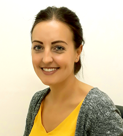
Michelle Dent discusses the process of transitioning into a new role and the pros and cons along the way.
An opportunity arose for a permanent, full time, band 7 specialist role in the medical retina (MR) team in the Newcastle Eye Centre (NEC) in December 2016. The position was open to nurses, optometrists and orthoptists. The role was for the clinician to be trained to treat patients in the MR clinics with intravitreal anti-vascular endothelial growth factor (anti-VEGF) injections and to make clinical decisions regarding patient management.
The relatively recent introduction of non-medical healthcare practitioners into this role is in response to the exponential growth in numbers of patients requiring the service, supported by the Royal College of Ophthalmologists (RCO) and the National Institute for Health and Care Excellence (NICE). The British and Irish Orthoptic Society (BIOS) have published standards of practice and guidelines specifically for orthoptists extending practice into this role. Some of the essential requirements were to have extensive post registration experience, good communication and interpersonal skills and to have the ability to work independently and across disciplines.

Michelle Dent.
The team already consisted of three nurse specialists undertaking this role, with a further three nurses more recently trained to carry out injections only. Following eight years working full time as an orthoptist, where I assess, diagnose and manage children and adults with vision and ocular motility problems, I applied for this role for further knowledge and experience in an area of ophthalmology that has always interested me. Following application and interview, I was able to accept the position on a part-time basis; working in MR for three days and in the orthoptic department for two days. The remaining two days of the MR post was filled by an optician who became my job share. I was assigned a mentor from the existing team and began to observe their daily work, taking on board a great deal of information about this completely new area of ophthalmology; observing the interpretation of optical coherence tomography (OCT) scans, slit lamp examinations, injections and how the clinics were organised and ran. At this early point I was able to appreciate the sheer volume of patients suffering from macular conditions and just what their journey through a MR appointment involved.
ANTT
Part of the initial training was to learn about aseptic non-touch technique (ANTT), the UK standardised aseptic technique to minimise the introduction of micro-organisms, which may occur during preparation, administration and delivery of an injection. I was already familiar with correct hand washing procedures from my experience in assessing orthoptic patients but ANTT was a new concept to me. I observed and then aided with the opening of a number of injection pre-packs onto the injection trolley, ensuring all required equipment was dropped onto the trolley from the appropriate height. I was then assessed and signed off as being competent in ANTT by a staff nurse who specialises in infection control.
Electronic patient records
One of the considerable differences in practice in MR is the use of electronic patient records on Medisoft, as this software has not yet been introduced into the orthoptic department. Initially this was challenging as I had no prior experience with the system and there seemed to be multiple windows to open and even more drop-down boxes to populate! However, regular use has allowed me to become much more competent and aware of its many advantages; the use of less paper, the fact that the records are legible, comprehensive, universal and eliminates problems that arise with regards to missing notes.
The only current disadvantage is the speed but this issue has been recognised. Medisoft also scores visual acuity as number of letters scored on the 85 letter ‘ETDRS’ logMAR chart, although can convert into the Snellen equivalent.
Having only ever worked with logMAR decimal scores, it took a little time to get used to this new method and to understand what level of vision a score related to.
Vision at each visit is automatically plotted onto a graph, which is a particularly good visual aid to show how vision has changed over time with progression of disease or with treatment.
This would be a valuable and time saving tool in the orthoptic department to see how a child’s vision is changing over the course of amblyopia treatment; to save filling in a paper graph by hand, as is done in current practice.
Developing an injection technique
Having observed multiple injections, I was then trained in the wet lab, where I practised the whole procedure on a model eye under supervision of a consultant ophthalmologist. Once confident enough in my ability, I began injecting patients under supervision. Following one hundred observed injections, twenty of those by a consultant, I was signed off as being competent and able to inject independently. Although an approved, generic protocol for injecting is always followed, techniques between clinicians vary somewhat and I have found myself adopting a mixture of them to come up with a technique that works for me, adapting it along the way following feedback from patients and colleagues.
Some of my own preferences include setting the bed to a slightly reclined position where possible, as I find the drape can sag in front of the eye if sitting upright, which can restrict my view of the eye. I have found that an infection control approved, wipe-down pillow sufficiently supports the patient’s neck for maximum comfort.
Once a drop of oxybuprocaine has been instilled to start the topical anaesthesia, I prefer the patient to keep their eyes closed. A drop of povidone-iodine is inserted into the lower fornix cul-de-sac and some onto the lashes, during which time I check the expiry date on the drug, apply an apron, undergo surgical disinfection and put on sterile gloves. This allows the recommended three minutes of contact time for the iodine. I personally prefer the trolley to be positioned on the patient’s right-hand side, regardless of which eye is undergoing the procedure.
Once chlorhexidine 0.1% aqueous solution has been used to clean the periocular skin, I then measure the prescribed drug to 0.05ml in the syringe (and draw up in cases of aflibercept) so as to allow the chlorhexidine solution at least thirty seconds of contact time. I have found that gently drying the face with gauze from the trolley ensures the drape sticks sufficiently.
Once I have inserted the speculum I have come to adopt the cotton swab tamponade technique, whereby a sterile swab is soaked in the anaesthetic and held on the site of injection for a few seconds.
This is following patient feedback that this technique allows for a more comfortable experience. Another drop of iodine is instilled onto the site of injection and given thirty seconds of contact time, which I utilise to carry out repeat checks of the eye and drug.
Following the injection and removal of the needle I have found placing a sterile cotton swab immediately over the injection site prevents reflux.
Following an antibiotic drop and ensuring the patient can count fingers (to ensure the central retinal artery is perfused), the speculum and drape are removed and the mark on the patient’s forehead wiped off; I always ensure this is after the procedure is complete to minimise risk of error throughout. I issue any unused sterile gauze to the patient at this point to gently dab around the eye or cheek if required, ensuring any old tissues are discarded.
Advice is given after every injection to refrain from touching or rubbing the eye, as well as recommendations for the immediate future such as to avoid swimming and eye make-up. It is ensured that the patient has their follow-up visit arranged and that they have a NEC contact card with emergency phone numbers on, should any problems arise.
New skills and terminology
My working week includes regular clinic sessions, where I analyse the results of different test results and combine this with a discussion with the patient to come up with a treatment decision. The opportunity to assess patients in clinic was one of the fundamental reasons that this post had initially appealed to me, as I was very keen to have a sound understanding of the conditions I was treating and to have an involvement in decisions.
This involves the interpretation of OCT images, which I had only briefly touched upon during my orthoptic studies at university but did not have any clinical experience with. I started to become familiar with some of the terms commonly used to describe OCT scans; intraretinal fluid, subretinal fluid, cysts, pigment epithelial detachments, retinal angiomatous proliferation lesions etc. It took time to learn what each of these meant, as well as their associated abbreviations!
I now feel comfortable with reviewing, describing and comparing OCT images to previous visits to identify and monitor signs of activity in macular conditions. I am able to carry out OCT scans myself using the Topcon 2000 OCT and the Heidelberg Spectralis machines, which can be a useful skill in busy outreach clinics to assure a constant patient flow.
Fundus examination using the slit lamp also contributes significantly to the clinical decision. Again, this is a machine I had little experience with and certainly no recent experience. At first, I practised finding the fundus on just about everyone I came into contact with, patients and colleagues alike! In time I became more efficient and more accustomed to recognising what was normal and what was not. In the early stages I found the most challenging aspect to be exploring different parts of the fundus by moving the Volk lens and asking the patient to look into certain positions of gaze, all while remembering the image is inverted and reversed, with different levels of magnification depending on strength of Volk lens used. With practise I became more competent with focusing accurately, adjusting brightness and slit width accordingly and ensuring I was achieving a stereoscopic view.
I started being able to identify and describe signs of macular conditions, such as drusen, haemorrhages, lesions and disciform scarring and as my understanding of the anatomy of the retina advanced further, this helped to identify the location of these abnormalities. There have been occasions where patients are unable to manoeuvre to the slit lamp, which has allowed me to practise headset binocular indirect ophthalmoscopy.
With increasing experience, I am picking up other useful skills on the slit lamp, such as Goldmann Applanation Tonometry (GAT) to measure intraocular pressures. I hope to continue improving my technique and expanding my skill set, in particular assessing patients without topical mydriasis, which can currently prove challenging.
Working in a new area of ophthalmology has brought with it a different category of patients. This does not only relate to the obvious difference in average age group but the background, level of support required, expectations and way in which symptoms can impact everyday life can vary greatly.
Despite this, many skills acquired in my two roles are transferable. Adults attending the orthoptic department can frequently also suffer from macular conditions and I am now able to give much more comprehensive information and advice, as well as having a greater understanding of their situation. For example, it may be less likely that a patient will have the ability to fuse with a Fresnel prism if their central vision is compromised.
As I believe having this knowledge can be beneficial to patients I am delivering a teaching session to orthoptic colleagues on macular conditions and their potential impact.
I will be providing Medisoft training once introduced into the orthoptic department and have also recently provided training to nurses in the wider clinics on strabismus and paediatric vision assessment. I am able to measure intraocular pressures using the handheld tonometer, which can be beneficial in orthoptic clinics where the nurses are occupied and there might otherwise be a delay for the patient.
There have been instances in macular clinics whereby patients have reported diplopia and a cover test establishes whether an onward referral to the orthoptic team would be beneficial. In some cases this diplopia is monocular in nature, due to distortion caused by macular disease.
Common challenges
I have been faced with numerous challenging experiences so far.
Understandably, given the nature of the procedure, patients can be extremely apprehensive and can struggle to remain still during the injection. There has been one incidence of abandoning the procedure due to patient anxiety. In this case the patient agreed to return one week later at which time they were considerably calmer.
With an ever-ageing population comes increasing patient numbers and a high demand on the service, resulting in busy clinics. Patients often require multiple tests at each visit to monitor their condition and time delays and bottle necks are inevitable.
Patients or family members can often be frustrated with the amount of time they have waited for their appointment or injection. Keeping patients informed of delays and giving forewarning when follow-up appointments may involve further tests is of utmost importance.
Other common challenges include patients who require injections to be administered while in wheelchairs, those who require an interpreter and it can sometimes be particularly difficult to insert the speculum in patients who find this part of the procedure extremely uncomfortable.
Given the average age of the patient in the MR clinic, it is not unusual to meet patients who have recently lost partners and this can be difficult. The Mental Capacity Act (MCA) 2005 and its accompanying Code of Practice is always at the forefront of my mind when patients are making treatment decisions. I have a dementia patient with a lack of mental capacity for which a lasting power of attorney is in place for a family member to make treatment decisions. I have also had a recent experience of an elderly gentleman declining treatment and accepting that vision is likely to deteriorate significantly.
Understanding when symptoms are related to other conditions is important, as is an awareness of when to refer on to another specialist, for example for cataract surgery. Prognosis can often be guarded and ensuring realistic expectations is vital.
Deaf patients relying on lip reading also require some forward thinking in the injection room, since face masks are worn throughout the procedure!
Support and education
Regular teaching sessions are provided in the NEC by a consultant ophthalmologist to support staff undertaking this extended role.
Opportunities to attend study days and workshops have allowed me expand on my knowledge of macular disease, as well as in wider fields such as national guidance for treatment, current research and potential future treatments. Consent training means I am able to consent / re-consent patients where necessary and attendance at the department’s regular fluorescein angiography meetings is giving me a greater insight into understanding and interpreting these tests. The orthoptic department regularly has undergraduate orthoptic students visiting on placement and I often provide a session to allow them to shadow the MR clinics and injections, giving them an insight into an orthoptic extended role that may become an option for them in their future careers.
I have now been in the post for one year and have carried out 1,226 injections to date. I believe this extended role has been positive for the NEC as a whole; I have become part of the wider multidisciplinary team and have promoted integration and sharing of information between departments.
My ophthalmology knowledge has expanded hugely, although I still have a lot to learn.
The position has undeniably opened my eyes as to just how many patients are seen daily within the NEC. I aim to continue improving and expanding my skill set and knowledge of macular conditions. I would like to conduct some of my own research into different injection techniques and their associated subjective pain scores and amount of time taken. I look forward to welcoming new ways of working, such as the possible introduction of the ‘intravitreal assistant’ device into the injection rooms and the introduction of virtual clinics. It has been a journey I have very much enjoyed and it is rewarding to know that I am helping to ease pressure on a busy service.
Even more rewarding is the difference that treatment can make to quality of life for some patients and they will often express their gratitude for the service.
COMMENTS ARE WELCOME


