This article has been verified for CPD. Click the button below to answer a few
short questions and download a form to be included in your CPD folder.

A comprehensive study evaluating the clinical indications, visual outcomes and complication rates of iris-claw intraocular lens (IOL) implantation in aphakic and complex IOL cases.
Loss of the capsule and/or zonules can result from various reasons, including complicated cataract surgery, trauma and ocular pathologies, such as pseudoexfoliation (PXF) syndrome, Marfan syndrome and lens coloboma. The correction of aphakia, dislocated or subluxated lens with inadequate support for the placement of a standard posterior chamber IOL (PCIOL) in the capsular bag or in the ciliary sulcus is challenging for surgeons [1].
Since the 1980s, four-point fixation flexible open-loop angle-supported anterior chamber IOL (ACIOL) has been used for lens implantation in eyes without capsular support. Other alternative methods have been developed, such as iris-fixation with iris-sutured IOL and iris-claw IOL (ICIOL), scleral-fixation by suturing of PCIOL or intrascleral haptic fixation of PCIOL [2]. Artisan aphakic IOL (Ophtec BV), the first ICIOL without need of suture for correction of aphakia, was developed in 1978 [3]. And according to many studies, ICIOL implantation either in the anterior chamber or by retro-pupillary fixation became an effective and safe choice with relatively simple placement and better clinical outcomes compared with scleral-fixation IOL or ACIOL [4]. The purpose of this study was to evaluate the indications, postoperative visual acuity and incidence of complication after intraocular implantation of iris-claw aphakic IOL (artisan IOL).
"Artisan IOL implantation offers a valuable solution in managing aphakia and complex secondary IOL cases"
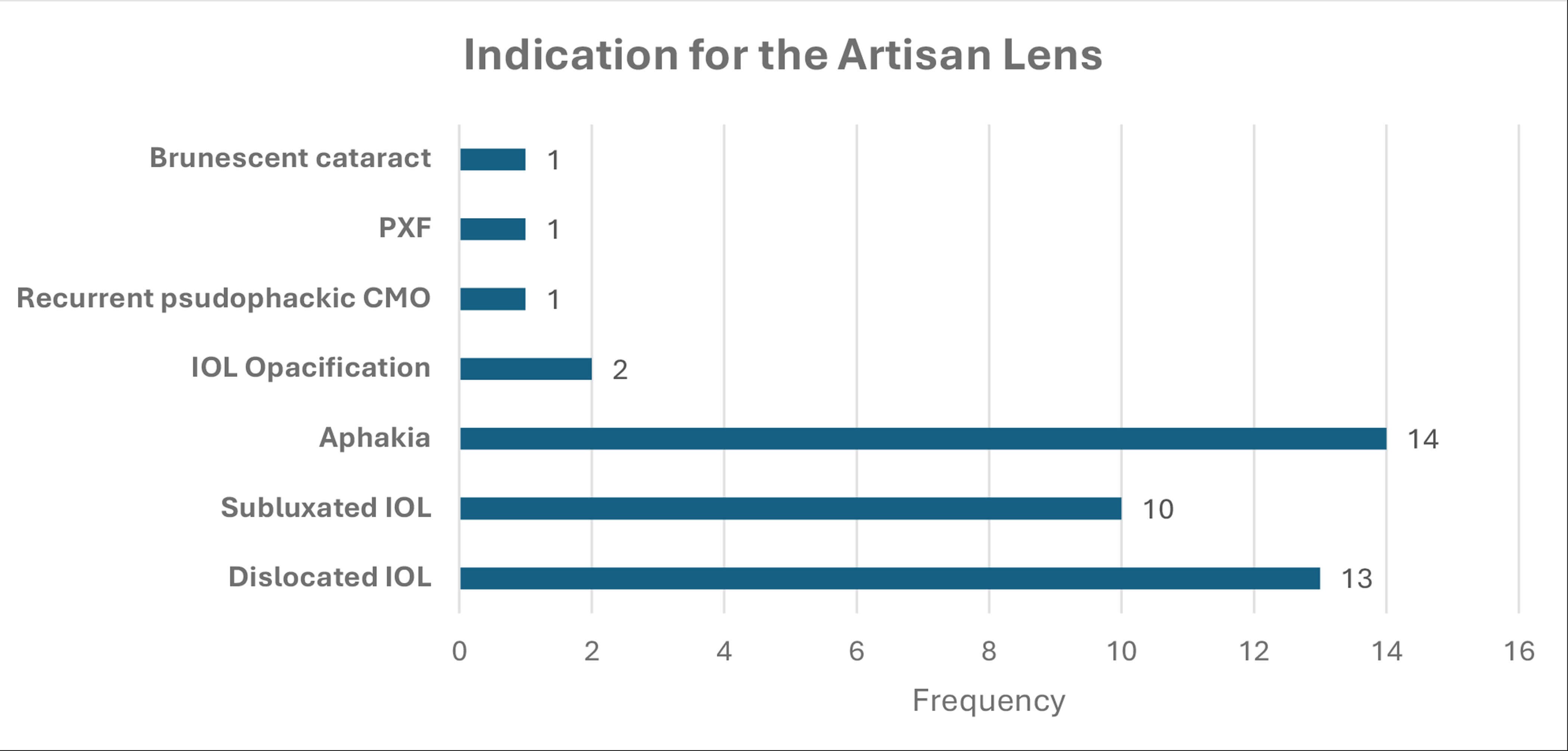
Figure 1: Frequency and percent of indication for the artisan lens.
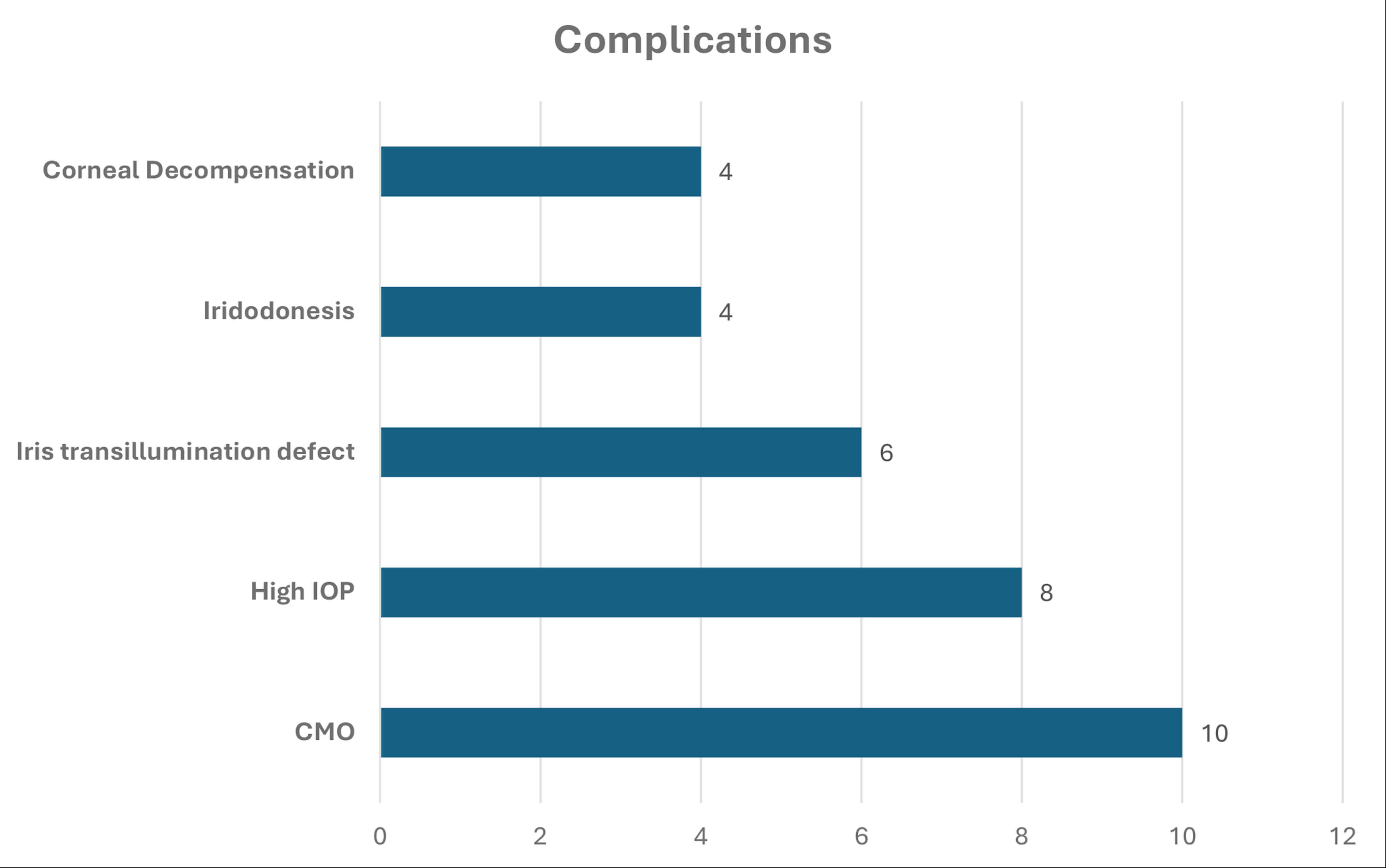
Figure 2: Frequency of complications.
Method and sample
Our method was a descriptive hospital-based study of 42 eyes (39 patients). Demographics, surgical details, complications and visual acuity are described. The study is a total coverage for all the patients that had their artisan IOL implantation surgeries in the period between April 2022 to January 2024 at Cork University Hospital/South Infirmary Victoria University Hospital. For the data collection, a closed-ended checklist was used, which contained:
- Detailed questions about the participants age and gender (demographic features)
- Indication of the artisan IOL implantations
- Position of the implanted lens
- Whether a surgical peripheral iridotomy (PI) was done or not
- Any preexisting ocular comorbidities
- Visual acuity (VA)
- Best corrected visual acuity (BCVA), and
- Incidence of complications.
The results of statistical analysis were performed using Statistical Package for the Social Sciences (SPSS) (Version 26) and Microsoft Excel and Word (2016). Descriptive statistics are presented in the form of frequency tables with percentages and graphical representations. Estimates of these percentages within the audit population were done through (95%) confidence interval. And for the hypotheses test, Chi square is presented with relevant cross tabulations. The P value of 0.05 or less is considered statistically significant.
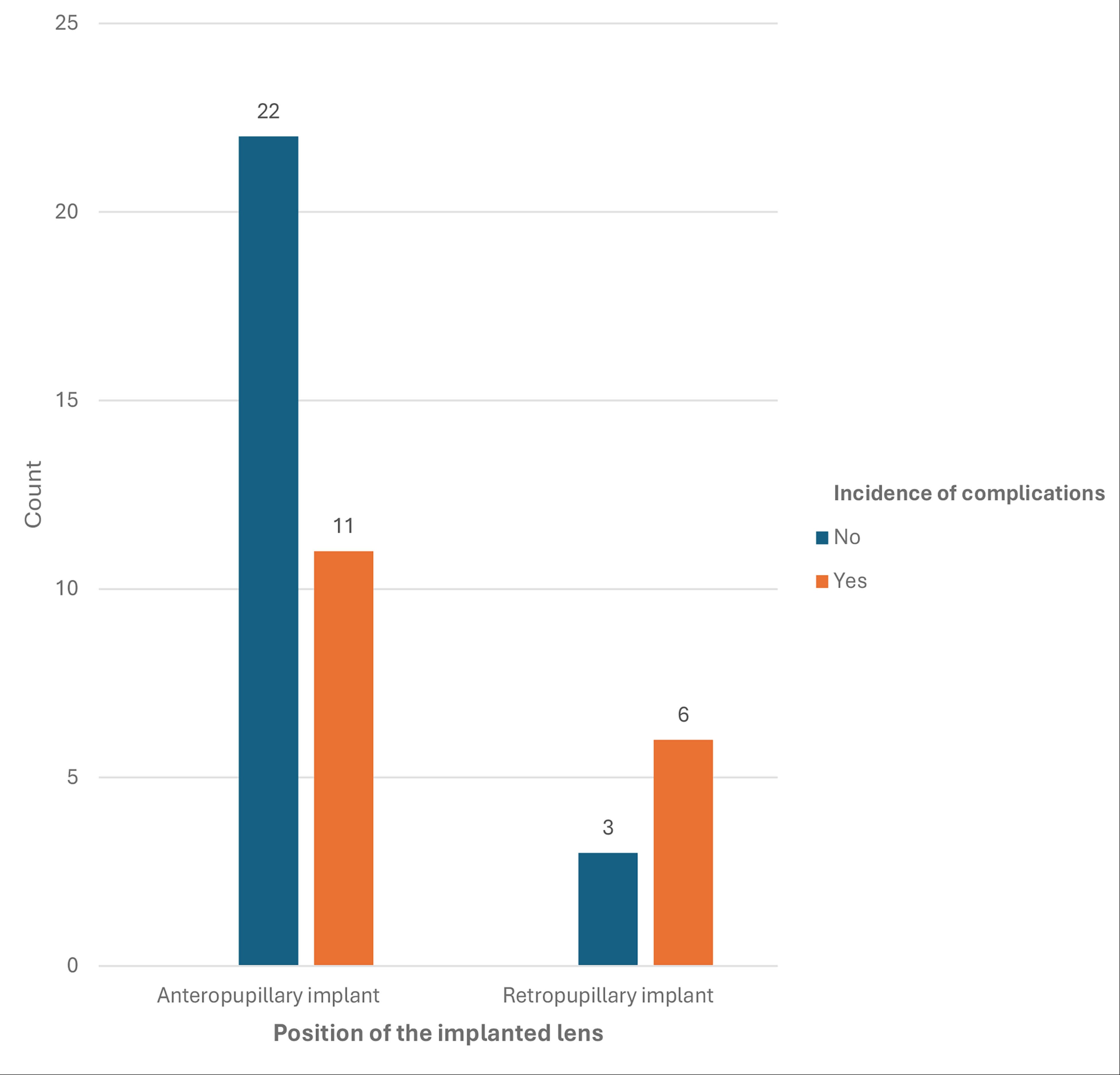
Figure 3: Association between position of the implanted lens and incidence of complications.
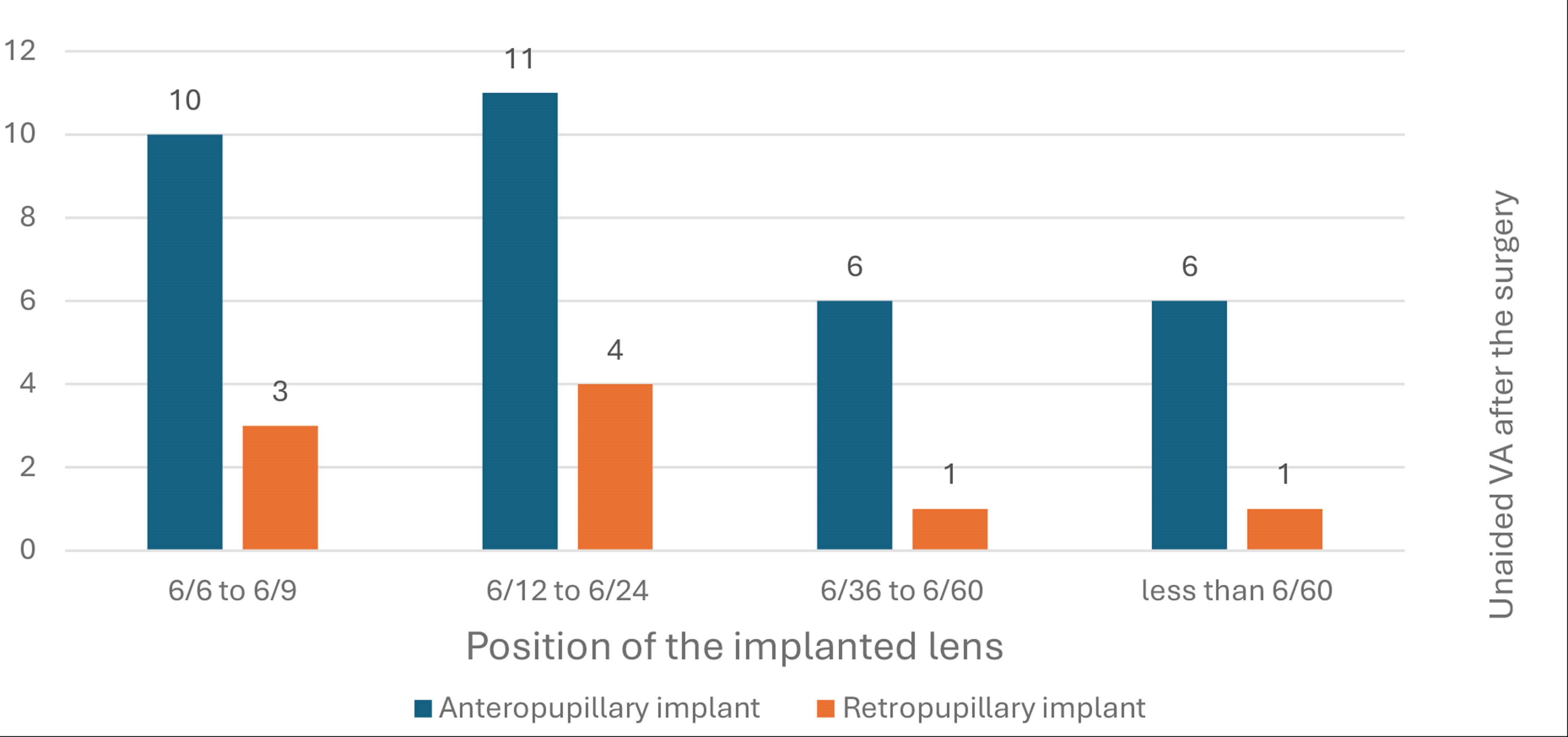
Figure 4: Association between unaided VA after the surgery and position of the implanted lens.
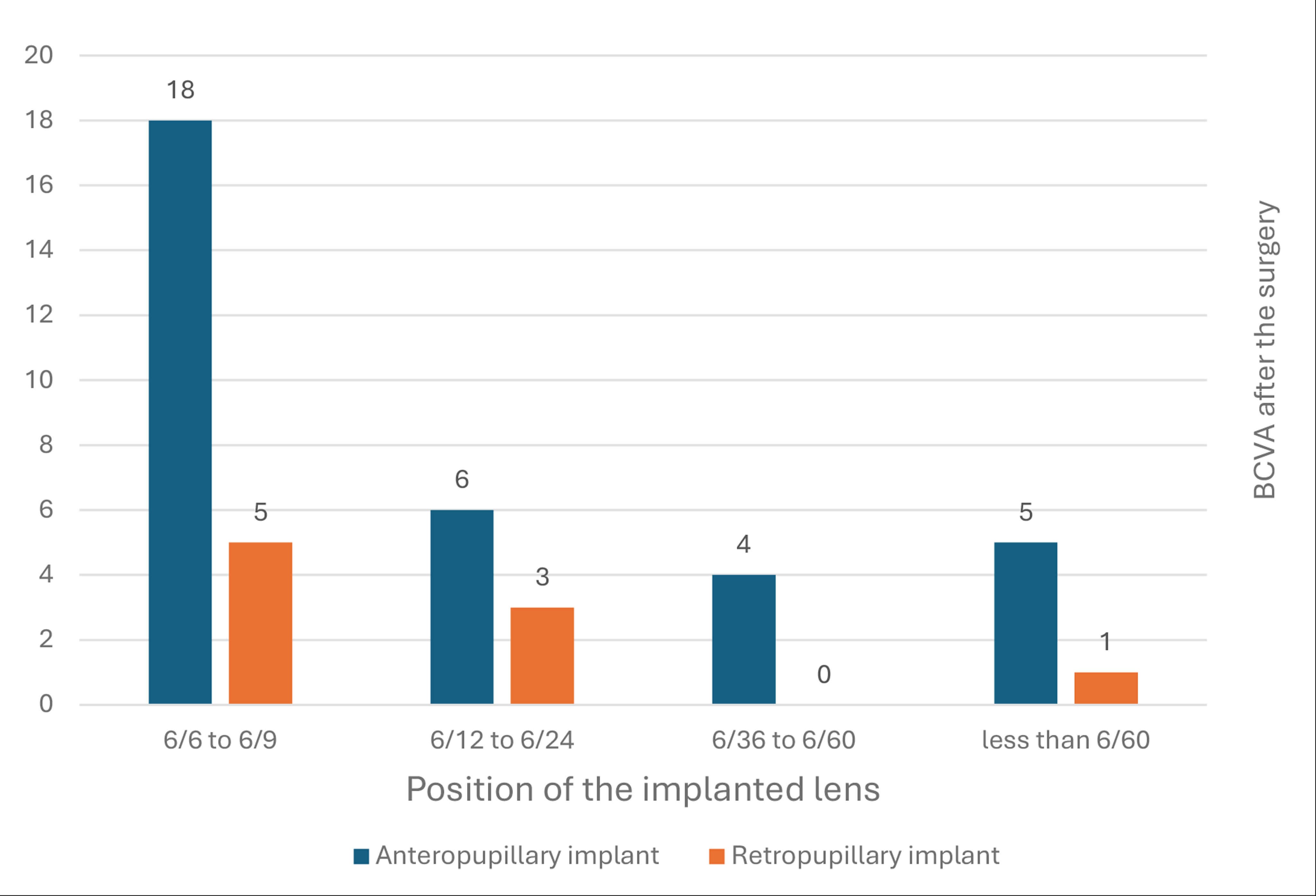
Figure 5: Association between BCVA after the surgery and position of the implanted lens.
Results
The mean age of the patients was 66.31 years old; 50% of the patients were males and 50% were females and this indicates that there is no specific age or gender for artisan IOL implantation indications. Most of the indications for artisan IOL implantation were aphakia (33.3%) and most of those aphakic eyes were due to complicated cataract surgeries, post-vitrectomy or ocular trauma surgeries. The second most common indications were dislocated IOL (31%) and subluxated IOL (23.8%) and most of those patients had Marfan’s syndrome, PXF syndrome or PXF glaucoma. The rest of the indications were due to explant of opacified IOL with the capsular complex, recurrent pseudophakic cystoid macular oedema (CMO) and Brunescent cataract. Regarding the position of the implanted lens, 78.6% were antero-pupillary and 21.4% of them were retro-pupillary implant.
According to these results, we found out there’s no association between position of the implanted lens and incidence of complications. However, there’s an association between position of the implanted lens and having high intraocular pressure (IOP) as a complication – a higher percentage of patients with retro-pupillary implant developed high IOP. Ninety-point-five percent of the patients had preexisting ocular comorbidities and 50% of those comorbidities were previous retinal detachment repair surgeries – most of them underwent cataract phacoemulsification without IOL implantation at the same time or soon after the retinal surgeries. Eighty-one percent of those patients had surgical PI done during the surgeries. The importance of surgical PI is to lower the risk of developing high IOP after the surgery. Forty-point-five percent of the patients developed complications, and most of them were CMO and high IOP; the rest differ between iris transillumination defects, iridodonesis and corneal decompensation. The VA was 6/6 to 6/9 in 31% of the patients, 6/12 to 6/24 in 35.7% of the patients, 6/36 to 6/60 in 16.7% of the patients, and less than 6/60 in 16.7% of the patients. And the BCVA was 6/6 to 6/9 in 54.8% of the patients, 6/12 to 6/24 in 21.4% of the patients, 6/36 to 6/60 in 9.5% of the patients, and less than 6/60 in 14.3% of the patients. We found that there’s no association between the VA and BCVA after the surgery and presence of comorbidities, as well as between the VA and BCVA and the position of the lens.
Discussion
The findings of this study further emphasise the growing body of evidence supporting the safety and effectiveness of ICIOL implantation for the management of aphakia and complex secondary IOL situations when capsular support is inadequate.
In our study, aphakia following complicated cataract surgery and intraocular lens dislocation were the most common indications, which is aligning with published literature describing similar case profiles [1-4]. Our study demonstrated favourable visual outcomes, with more than half of the patients achieving a BCVA of 6/6–6/9 postoperatively. This is consistent with Liang, et al.’s systematic review, which reported good postoperative visual outcomes in both anterior and retro-pupillary artisan IOL fixation groups [1]. These assure the reliability of the artisan IOL as a visually effective solution for such cases.
Complication rates in our cohort were acceptable for a high-risk surgical population, with CMO and elevated IOP being the most frequent postoperative events. In comparison with findings that were reported by Galvis, et al., who noted CMO as one of the common complications following retro-pupillary ICIOL implantation, particularly in eyes with previous retinal pathology [3]. Jing, et al.’s meta-analysis further supports the observation that postoperative inflammation related complications occur at similar rates between scleral fixated and iris claw lenses [4].
Of particular interest in our study was the statistically significant association between retro-pupillary implantation and postoperative elevation of IOP. Liang, et al. likewise observed that retro-pupillary implantation is altering aqueous flow dynamics [1]. The majority of eyes in our study underwent surgical peripheral iridotomy if they had not had it for any other reason before the surgery, which helps in avoiding the risk of pupillary block, positively affecting postoperative IOP.
Our findings also reflect those of Shen, et al., who highlighted that iris claw IOLs, whether anterior or retro-pupillary, demonstrate stable long-term fixation with low rates of lens displacements and minimal endothelial cell loss when performed with correct technique and appropriate case selection [2]. The absence of significant differences in corneal decompensation between the two positions in our study reinforces this safety profile.
Another important observation was the lack of association between preexisting ocular comorbidities and final BCVA. Despite many patients having undergone previous retinal detachment repair, postoperative vision remained favourable. This echoes the results of Galvis, et al., who found that iris claw implantation maintained good functional outcomes even in surgically complex eyes [3].
These similarities suggest that artisan IOL implantation remains a robust option even when performed in eyes with substantial comorbidity. Overall, the outcomes of this audit confirm that the artisan IOL – whether placed anteriorly or retro pupillary – offers a dependable solution for aphakia and IOL instability, with a predictable visual prognosis and complication profile comparable to international experience. Given its relative ease of implantation, reduced risk of suture-related complications and strong visual outcomes, the iris claw lens remains an attractive choice in tertiary surgical settings. Future prospective studies with larger sample sizes and longer follow-up intervals would be valuable in clarifying the long-term endothelial safety and optimising the decision-making around implantation position.
Conclusion
This study reveals that there is no specific age or gender for artisan lens implantation indications. Most of the indications for artisan lens implantation were aphakia then dislocated and subluxated IOL. Artisan IOL implantation offers a valuable solution in managing aphakia and complex secondary IOL cases. Most of the preexisting ocular comorbidities were previous retinal detachment repair surgeries. And most of the patients had surgical PI done. Less than half of the patients developed complications after the surgery and most of them were CMO and high IOP and with a lower incidence they developed iris transillumination defects, iridodonesis and corneal decompensation. Despite higher IOP in the retro-pupillary group, visual outcomes remained favourable across the board. Regarding the visual outcomes, we found there to be no association between the VA and BCVA after the surgery, the presence of comorbidities or the position of the implanted lens. These results support broader use in tertiary care settings.
References
1. Liang IC, Chang YH, Hernández Martínez A, Hung CF. Iris-claw intraocular lens: anterior chamber or retropupillary implantation? A systematic review and meta-analysis. Medicina (Kaunas) 2021;57(8):785.
2. Shen JF, Deng S, Hammersmith KM, et al. Intraocular lens implantation in the absence of zonular support: an outcomes and safety update: a report by the American Academy of Ophthalmology. Ophthalmology 2020;127(9):1234–58.
3. Galvis V, Tello A, Carreño NI, Revelo ML. Retropupillary iris-claw intraocular lens in aphakic eyes. J Cataract Refract Surg 2013;39(6):970–1.
4. Jing W, Guanlu L, Qianyin Z, et al. Iris-claw intraocular lens and scleral-fixated posterior chamber intraocular lens implantations in correcting aphakia: a meta-analysis. Investig Ophthalmol Vis Sci 2017;58(9):3530–6.
Declaration of competing interests: None declared.
Acknowledgements: This study was awarded the John Blake Medal for ‘Best Scientific Ophthalmology Research Paper’ in Ireland, November 2024, in recognition of its contribution to ophthalmic surgical outcomes research. We express our gratitude to the medical records centre at Cork University Hospital/South Infirmary Victoria University Hospital for their assistance in conducting this study. We are highly appreciative to all the surgeons who conducted the artisan IOL implantation and all the medical staff who provided patient care.